1UAS | pdb_00001uas

Crystal structure of rice alpha-galactosidase


Experimental Data Snapshot

  • Method: X-RAY DIFFRACTION
  • Resolution: 1.50 Å
  • R-Value Free: 
    0.178 (Depositor), 0.180 (DCC) 
  • R-Value Work: 
    0.160 (Depositor), 0.160 (DCC) 
  • R-Value Observed: 
    0.160 (Depositor) 

wwPDB Validation   3D Report Full Report


Ligand Structure Quality Assessment 

Created with Raphaël 2.3.0Worse 01 BetterLigand structure goodness of fit to experimental dataBest fitted GLAClick on this verticalbar to view details

This is version 1.5 of the entry. See complete history


Literature

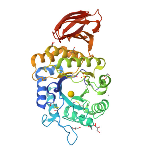
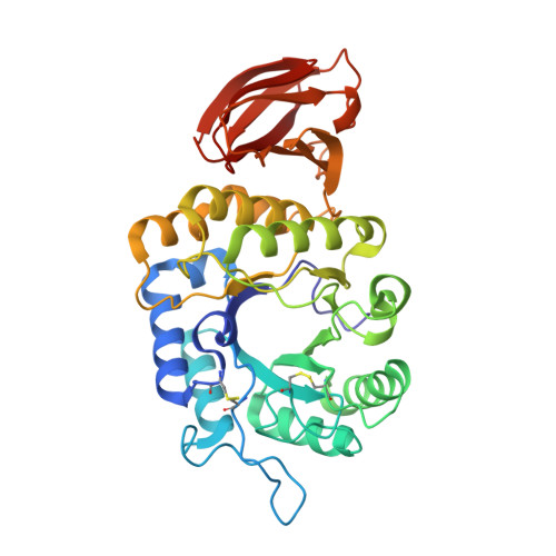
Crystal structure of rice alpha-galactosidase complexed with D-galactose

Fujimoto, Z.Kaneko, S.Momma, M.Kobayashi, H.Mizuno, H.

(2003) J Biological Chem 278: 20313-20318

  • DOI: https://doi.org/10.1074/jbc.M302292200
  • Primary Citation of Related Structures:  
    1UAS

  • PubMed Abstract: 

    alpha-Galactosidases catalyze the hydrolysis of alpha-1,6-linked galactosyl residues from galacto-oligosaccharides and polymeric galacto-(gluco)mannans. The crystal structure of rice alpha-galactosidase has been determined at 1.5A resolution using the multiple isomorphous replacement method. The structure consisted of a catalytic domain and a C-terminal domain and was essentially the same as that of alpha-N-acetylgalactosaminidase, which is the same member of glycosyl hydrolase family 27. The catalytic domain had a (beta/alpha)8-barrel structure, and the C-terminal domain was made up of eight beta-strands containing a Greek key motif. The structure was solved as a complex with d-galactose, providing a mode of substrate binding in detail. The d-galactose molecule was found bound in the active site pocket on the C-terminal side of the central beta-barrel of the catalytic domain. The d-galactose molecule consisted of a mixture of two anomers present in a ratio equal to their natural abundance. Structural comparisons of rice alpha-galactosidase with chicken alpha-N-acetylgalactosaminidase provided further understanding of the substrate recognition mechanism in these enzymes.


  • Organizational Affiliation

    Department of Biochemistry, National Institute of Agrobiological Sciences, Tsukuba, Ibaraki 305-8602, Japan.


Macromolecules
Find similar proteins by:  (by identity cutoff)  |  3D Structure
Entity ID: 1
MoleculeChains Sequence LengthOrganismDetailsImage
alpha-galactosidase362Oryza sativaMutation(s): 0 
EC: 3.2.1.22
UniProt
Find proteins for Q9FXT4 (Oryza sativa subsp. japonica)
Explore Q9FXT4 
Go to UniProtKB:  Q9FXT4
Entity Groups  
Sequence Clusters30% Identity50% Identity70% Identity90% Identity95% Identity100% Identity
UniProt GroupQ9FXT4
Sequence Annotations
Expand
  • Reference Sequence
Small Molecules
Ligands 4 Unique
IDChains Name / Formula / InChI Key2D Diagram3D Interactions
PT
Query on PT

Download Ideal Coordinates CCD File 
D [auth A]PLATINUM (II) ION
Pt
HRGDZIGMBDGFTC-UHFFFAOYSA-N
GLA
Query on GLA

Download Ideal Coordinates CCD File 
B [auth A]alpha-D-galactopyranose
C6 H12 O6
WQZGKKKJIJFFOK-PHYPRBDBSA-N
SO4
Query on SO4

Download Ideal Coordinates CCD File 
C [auth A]SULFATE ION
O4 S
QAOWNCQODCNURD-UHFFFAOYSA-L
GOL
Query on GOL

Download Ideal Coordinates CCD File 
E [auth A]
F [auth A]
G [auth A]
H [auth A]
I [auth A]
GLYCEROL
C3 H8 O3
PEDCQBHIVMGVHV-UHFFFAOYSA-N
Experimental Data & Validation

Experimental Data

  • Method: X-RAY DIFFRACTION
  • Resolution: 1.50 Å
  • R-Value Free:  0.178 (Depositor), 0.180 (DCC) 
  • R-Value Work:  0.160 (Depositor), 0.160 (DCC) 
  • R-Value Observed: 0.160 (Depositor) 
Space Group: P 21 21 21
Unit Cell:
Length ( Å )Angle ( ˚ )
a = 63.656α = 90
b = 71.42β = 90
c = 86.238γ = 90
Software Package:
Software NamePurpose
CNSrefinement
HKL-2000data reduction
SCALEPACKdata scaling
MLPHAREphasing

Structure Validation

View Full Validation Report


Ligand Structure Quality Assessment 

Created with Raphaël 2.3.0Worse 01 BetterLigand structure goodness of fit to experimental dataBest fitted GLAClick on this verticalbar to view details

Entry History 

Deposition Data

Revision History  (Full details and data files)

  • Version 1.0: 2003-07-01
    Type: Initial release
  • Version 1.1: 2008-04-27
    Changes: Version format compliance
  • Version 1.2: 2011-07-13
    Changes: Non-polymer description, Version format compliance
  • Version 1.3: 2020-07-29
    Type: Remediation
    Reason: Carbohydrate remediation
    Changes: Advisory, Data collection, Derived calculations, Structure summary
  • Version 1.4: 2023-12-27
    Changes: Data collection, Database references, Structure summary
  • Version 1.5: 2024-10-30
    Changes: Structure summary