1Z5L | pdb_00001z5l

Structure of a highly potent short-chain galactosyl ceramide agonist bound to CD1D


Experimental Data Snapshot

  • Method: X-RAY DIFFRACTION
  • Resolution: 2.20 Å
  • R-Value Free: 
    0.294 (Depositor), 0.290 (DCC) 
  • R-Value Work: 
    0.242 (Depositor), 0.240 (DCC) 
  • R-Value Observed: 
    0.243 (Depositor) 

Starting Model: experimental
View more details

wwPDB Validation   3D Report Full Report


Ligand Structure Quality Assessment 

Created with Raphaël 2.3.0Worse 01 BetterLigand structure goodness of fit to experimental dataBest fitted PBSClick on this verticalbar to view detailsBest fitted R16Click on this verticalbar to view detailsBest fitted NAGClick on this verticalbar to view details

This is version 2.2 of the entry. See complete history


Literature

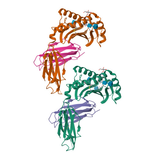
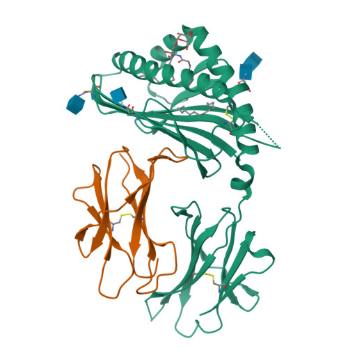
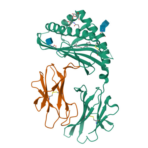
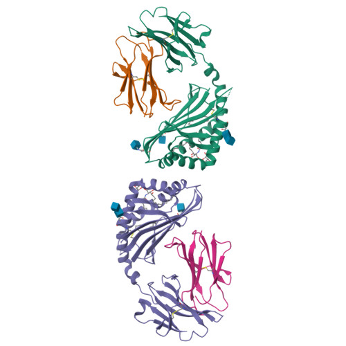
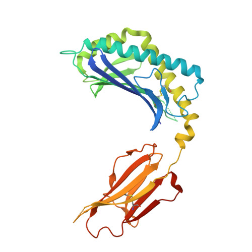
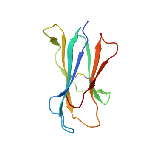
Structure and function of a potent agonist for the semi-invariant natural killer T cell receptor.

Zajonc, D.M.Cantu, C.Mattner, J.Zhou, D.Savage, P.B.Bendelac, A.Wilson, I.A.Teyton, L.

(2005) Nat Immunol 6: 810-818

  • DOI: https://doi.org/10.1038/ni1224
  • Primary Citation of Related Structures:  
    1Z5L

  • PubMed Abstract: 

    Natural killer T cells express a conserved, semi-invariant alphabeta T cell receptor that has specificity for self glycosphingolipids and microbial cell wall alpha-glycuronosylceramide antigens presented by CD1d molecules. Here we report the crystal structure of CD1d in complex with a short-chain synthetic variant of alpha-galactosylceramide at a resolution of 2.2 A. This structure elucidates the basis for the high specificity of these microbial ligands and explains the restriction of the alpha-linkage as a unique pathogen-specific pattern-recognition motif. Comparison of the binding of altered lipid ligands to CD1d and T cell receptors suggested that the differential T helper type 1-like and T helper type 2-like properties of natural killer T cells may originate largely from differences in their 'loading' in different cell types and hence in their tissue distribution in vivo.


  • Organizational Affiliation

    Department of Molecular Biology and The Scripps Research Institute, La Jolla, California 92037, USA.


Macromolecules
Find similar proteins by:  (by identity cutoff)  |  3D Structure
Entity ID: 1
MoleculeChains Sequence LengthOrganismDetailsImage
T-cell surface glycoprotein CD1d antigen
A, C
285Mus musculusMutation(s): 0 
UniProt & NIH Common Fund Data Resources
Find proteins for P11609 (Mus musculus)
Explore P11609 
Go to UniProtKB:  P11609
IMPC:  MGI:107674
Entity Groups  
Sequence Clusters30% Identity50% Identity70% Identity90% Identity95% Identity100% Identity
UniProt GroupP11609
Glycosylation
Glycosylation Sites: 3Go to GlyGen: P11609-1
Sequence Annotations
Expand
  • Reference Sequence
Find similar proteins by:  (by identity cutoff)  |  3D Structure
Entity ID: 2
MoleculeChains Sequence LengthOrganismDetailsImage
Beta-2-microglobulin
B, D
99Mus musculusMutation(s): 0 
UniProt & NIH Common Fund Data Resources
Find proteins for P01887 (Mus musculus)
Explore P01887 
Go to UniProtKB:  P01887
IMPC:  MGI:88127
Entity Groups  
Sequence Clusters30% Identity50% Identity70% Identity90% Identity95% Identity100% Identity
UniProt GroupP01887
Sequence Annotations
Expand
  • Reference Sequence
Oligosaccharides

Help

Entity ID: 3
MoleculeChains Length2D Diagram Glycosylation3D Interactions
2-acetamido-2-deoxy-beta-D-glucopyranose-(1-4)-2-acetamido-2-deoxy-beta-D-glucopyranose
E, F
2N-Glycosylation
Glycosylation Resources
GlyTouCan:  G42666HT
GlyCosmos:  G42666HT
GlyGen:  G42666HT
Small Molecules
Ligands 3 Unique
IDChains Name / Formula / InChI Key2D Diagram3D Interactions
PBS
Query on PBS

Download Ideal Coordinates CCD File 
I [auth A],
L [auth C]
(2S,3S,4R)-N-OCTANOYL-1-[(ALPHA-D-GALACTOPYRANOSYL)OXY]-2-AMINO-OCTADECANE-3,4-DIOL
C32 H63 N O9
PFXOKSFCNLTBGK-YFWOXBOYSA-N
R16
Query on R16

Download Ideal Coordinates CCD File 
J [auth A],
M [auth C]
HEXADECANE
C16 H34
DCAYPVUWAIABOU-UHFFFAOYSA-N
NAG
Query on NAG

Download Ideal Coordinates CCD File 
G [auth A],
H [auth A],
K [auth C]
2-acetamido-2-deoxy-beta-D-glucopyranose
C8 H15 N O6
OVRNDRQMDRJTHS-FMDGEEDCSA-N
Experimental Data & Validation

Experimental Data

  • Method: X-RAY DIFFRACTION
  • Resolution: 2.20 Å
  • R-Value Free:  0.294 (Depositor), 0.290 (DCC) 
  • R-Value Work:  0.242 (Depositor), 0.240 (DCC) 
  • R-Value Observed: 0.243 (Depositor) 
Space Group: P 1 21 1
Unit Cell:
Length ( Å )Angle ( ˚ )
a = 59.451α = 90
b = 77.045β = 107.63
c = 111.014γ = 90
Software Package:
Software NamePurpose
REFMACrefinement
HKL-2000data reduction
CCP4data scaling
MOLREPphasing

Structure Validation

View Full Validation Report


Ligand Structure Quality Assessment 

Created with Raphaël 2.3.0Worse 01 BetterLigand structure goodness of fit to experimental dataBest fitted PBSClick on this verticalbar to view detailsBest fitted R16Click on this verticalbar to view detailsBest fitted NAGClick on this verticalbar to view details

Entry History 

Deposition Data

Revision History  (Full details and data files)

  • Version 1.0: 2005-07-19
    Type: Initial release
  • Version 1.1: 2008-04-30
    Changes: Version format compliance
  • Version 1.2: 2011-07-13
    Changes: Advisory, Version format compliance
  • Version 2.0: 2020-07-29
    Type: Remediation
    Reason: Carbohydrate remediation
    Changes: Advisory, Atomic model, Data collection, Database references, Derived calculations, Structure summary
  • Version 2.1: 2023-08-23
    Changes: Advisory, Data collection, Database references, Refinement description, Structure summary
  • Version 2.2: 2024-10-30
    Changes: Structure summary