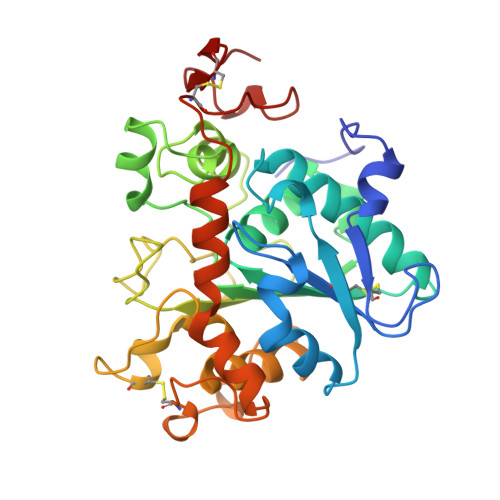
1TCC

THE SEQUENCE, CRYSTAL STRUCTURE DETERMINATION AND REFINEMENT OF TWO CRYSTAL FORMS OF LIPASE B FROM CANDIDA ANTARCTICA


Experimental Data Snapshot

  • Method: X-RAY DIFFRACTION
  • Resolution: 2.50 Å
  • R-Value Work: 0.196 
  • R-Value Observed: 0.196 

wwPDB Validation   3D Report Full Report


Ligand Structure Quality Assessment 


This is version 2.1 of the entry. See complete history


Literature

The sequence, crystal structure determination and refinement of two crystal forms of lipase B from Candida antarctica.

Uppenberg, J.Hansen, M.T.Patkar, S.Jones, T.A.

(1994) Structure 2: 293-308

  • DOI: https://doi.org/10.1016/s0969-2126(00)00031-9
  • Primary Citation of Related Structures:  
    1TCA, 1TCB, 1TCC

  • PubMed Abstract: 

    Lipases constitute a family of enzymes that hydrolyze triglycerides. They occur in many organisms and display a wide variety of substrate specificities. In recent years, much progress has been made towards explaining the mechanism of these enzymes and their ability to hydrolyze their substrates at an oil-water interface. We have determined the DNA and amino acid sequences for lipase B from the yeast Candida antarctica. The primary sequence has no significant homology to any other known lipase and deviates from the consensus sequence around the active site serine that is found in other lipases. We have determined the crystal structure of this enzyme using multiple isomorphous replacement methods for two crystal forms. Models for the orthorhombic and monoclinic crystal forms of the enzyme have been refined to 1.55 A and 2.1 A resolution, respectively. Lipase B is an alpha/beta type protein that has many features in common with previously determined lipase structures and other related enzymes. In the monoclinic crystal form, lipid-like molecules, most likely beta-octyl glucoside, can be seen close to the active site. The behaviour of these lipid molecules in the crystal structure has been studied at different pH values. The structure of Candida antarctica lipase B shows that the enzyme has a Ser-His-Asp catalytic triad in its active site. The structure appears to be in an 'open' conformation with a rather restricted entrance to the active site. We believe that this accounts for the substrate specificity and high degree of stereospecificity of this lipase.


  • Organizational Affiliation

    Department of Molecular Biology, Uppsala University, Sweden.


Macromolecules
Find similar proteins by:  (by identity cutoff)  |  3D Structure
Entity ID: 1
MoleculeChains Sequence LengthOrganismDetailsImage
LIPASE
A, B
317Moesziomyces antarcticusMutation(s): 0 
EC: 3.1.1.3
UniProt
Find proteins for P41365 (Pseudozyma antarctica)
Explore P41365 
Go to UniProtKB:  P41365
Entity Groups  
Sequence Clusters30% Identity50% Identity70% Identity90% Identity95% Identity100% Identity
UniProt GroupP41365
Glycosylation
Glycosylation Sites: 1
Sequence Annotations
Expand
  • Reference Sequence
Oligosaccharides

Help

Entity ID: 2
MoleculeChains Length2D Diagram Glycosylation3D Interactions
2-acetamido-2-deoxy-beta-D-glucopyranose-(1-4)-2-acetamido-2-deoxy-beta-D-glucopyranose
C
2N-Glycosylation
Glycosylation Resources
GlyTouCan:  G42666HT
GlyCosmos:  G42666HT
GlyGen:  G42666HT
Small Molecules
Ligands 1 Unique
IDChains Name / Formula / InChI Key2D Diagram3D Interactions
BOG
Query on BOG

Download Ideal Coordinates CCD File 
D [auth B]octyl beta-D-glucopyranoside
C14 H28 O6
HEGSGKPQLMEBJL-RKQHYHRCSA-N
Experimental Data & Validation

Experimental Data

  • Method: X-RAY DIFFRACTION
  • Resolution: 2.50 Å
  • R-Value Work: 0.196 
  • R-Value Observed: 0.196 
  • Space Group: P 1 21 1
Unit Cell:
Length ( Å )Angle ( ˚ )
a = 67α = 90
b = 50.5β = 100.1
c = 86.7γ = 90
Software Package:
Software NamePurpose
X-PLORmodel building
X-PLORrefinement
X-PLORphasing

Structure Validation

View Full Validation Report


Ligand Structure Quality Assessment 


Entry History 

Deposition Data

Revision History  (Full details and data files)

  • Version 1.0: 1994-05-31
    Type: Initial release
  • Version 1.1: 2008-03-03
    Changes: Version format compliance
  • Version 1.2: 2011-07-13
    Changes: Non-polymer description, Version format compliance
  • Version 1.3: 2017-11-29
    Changes: Derived calculations, Other
  • Version 2.0: 2020-07-29
    Type: Remediation
    Reason: Carbohydrate remediation
    Changes: Atomic model, Data collection, Derived calculations, Structure summary
  • Version 2.1: 2024-10-23
    Changes: Data collection, Database references, Structure summary