2AR8

The structure of tryptophan 7-halogenase (PrnA)suggests a mechanism for regioselective chlorination


Experimental Data Snapshot

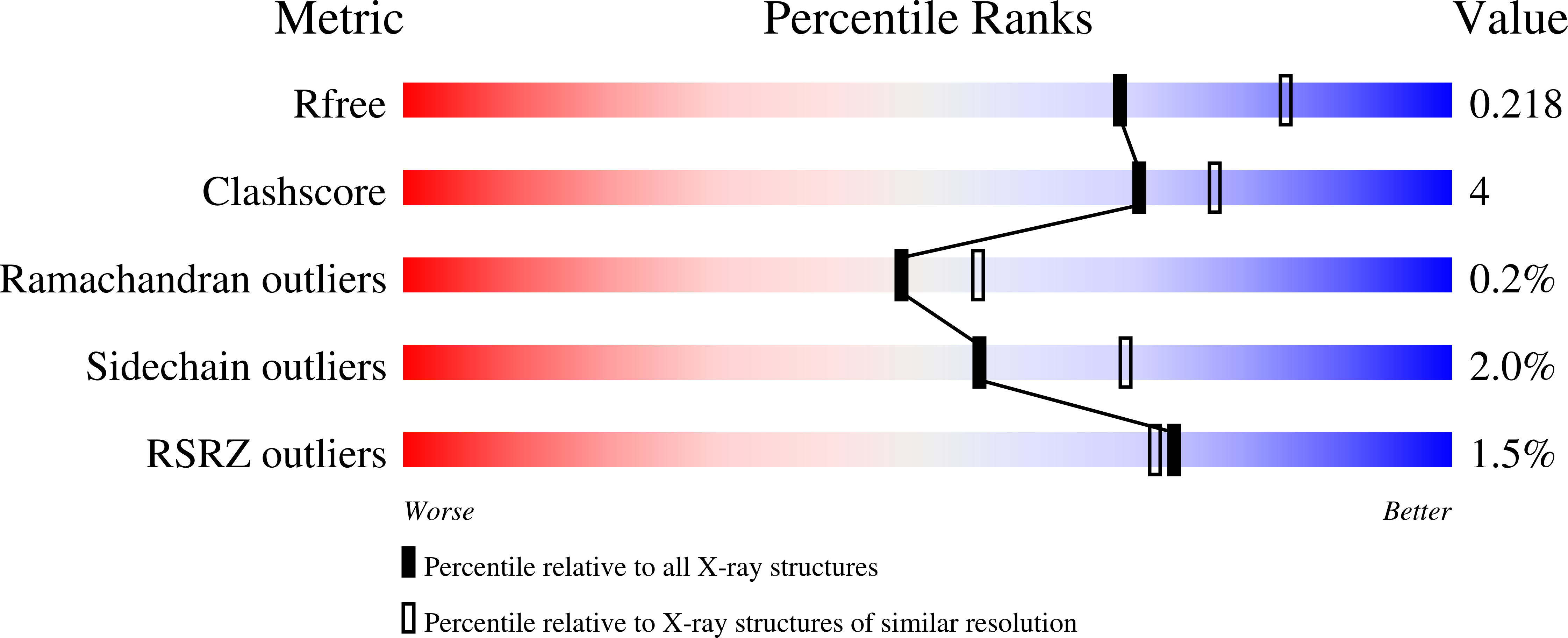
  • Method: X-RAY DIFFRACTION
  • Resolution: 2.20 Å
  • R-Value Free: 0.217 
  • R-Value Work: 0.172 
  • R-Value Observed: 0.173 

Starting Model: experimental
View more details

wwPDB Validation   3D Report Full Report


Ligand Structure Quality Assessment 


This is version 1.4 of the entry. See complete history


Literature

Tryptophan 7-halogenase (PrnA) structure suggests a mechanism for regioselective chlorination.

Dong, C.Flecks, S.Unversucht, S.Haupt, C.van Pee, K.H.Naismith, J.H.

(2005) Science 309: 2216-2219

  • DOI: https://doi.org/10.1126/science.1116510
  • Primary Citation of Related Structures:  
    2APG, 2AQJ, 2AR8, 2ARD

  • PubMed Abstract: 

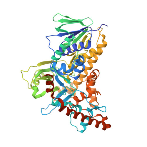
    Chlorinated natural products include vancomycin and cryptophycin A. Their biosynthesis involves regioselective chlorination by flavin-dependent halogenases. We report the structural characterization of tryptophan 7-halogenase (PrnA), which regioselectively chlorinates tryptophan. Tryptophan and flavin adenine dinucleotide (FAD) are separated by a 10 angstrom-long tunnel and bound by distinct enzyme modules. The FAD module is conserved in halogenases and is related to flavin-dependent monooxygenases. On the basis of biochemical studies, crystal structures, and by analogy with monooxygenases, we predict that FADH2 reacts with O2 to make peroxyflavin, which is decomposed by Cl-. The resulting HOCl is guided through the tunnel to tryptophan, where it is activated to participate in electrophilic aromatic substitution.


  • Organizational Affiliation

    Centre for Biomolecular Sciences, EaStchem, University of St. Andrews, St. Andrews KY16 9ST, UK.


Macromolecules
Find similar proteins by:  (by identity cutoff)  |  3D Structure
Entity ID: 1
MoleculeChains Sequence LengthOrganismDetailsImage
tryptophan halogenase PrnA538Pseudomonas fluorescensMutation(s): 0 
Gene Names: PrnA
EC: 1.14.19.9
UniProt
Find proteins for P95480 (Pseudomonas fluorescens)
Explore P95480 
Go to UniProtKB:  P95480
Entity Groups  
Sequence Clusters30% Identity50% Identity70% Identity90% Identity95% Identity100% Identity
UniProt GroupP95480
Sequence Annotations
Expand
  • Reference Sequence
Experimental Data & Validation

Experimental Data

  • Method: X-RAY DIFFRACTION
  • Resolution: 2.20 Å
  • R-Value Free: 0.217 
  • R-Value Work: 0.172 
  • R-Value Observed: 0.173 
  • Space Group: P 43 21 2
Unit Cell:
Length ( Å )Angle ( ˚ )
a = 67.128α = 90
b = 67.128β = 90
c = 275.702γ = 90
Software Package:
Software NamePurpose
SCALAdata scaling
REFMACrefinement
PDB_EXTRACTdata extraction
MOSFLMdata reduction
CCP4data scaling
MOLREPphasing

Structure Validation

View Full Validation Report


Ligand Structure Quality Assessment 


Entry History 

Deposition Data

Revision History  (Full details and data files)

  • Version 1.0: 2005-10-04
    Type: Initial release
  • Version 1.1: 2008-04-30
    Changes: Version format compliance
  • Version 1.2: 2011-07-13
    Changes: Advisory, Version format compliance
  • Version 1.3: 2023-08-23
    Changes: Data collection, Database references, Derived calculations, Refinement description
  • Version 1.4: 2023-11-15
    Changes: Data collection