2BFQ

MACRO DOMAINS ARE ADP-RIBOSE BINDING MOLECULES


Experimental Data Snapshot

  • Method: X-RAY DIFFRACTION
  • Resolution: 1.50 Å
  • R-Value Free: 0.233 
  • R-Value Work: 0.210 
  • R-Value Observed: 0.210 

Starting Model: experimental
View more details

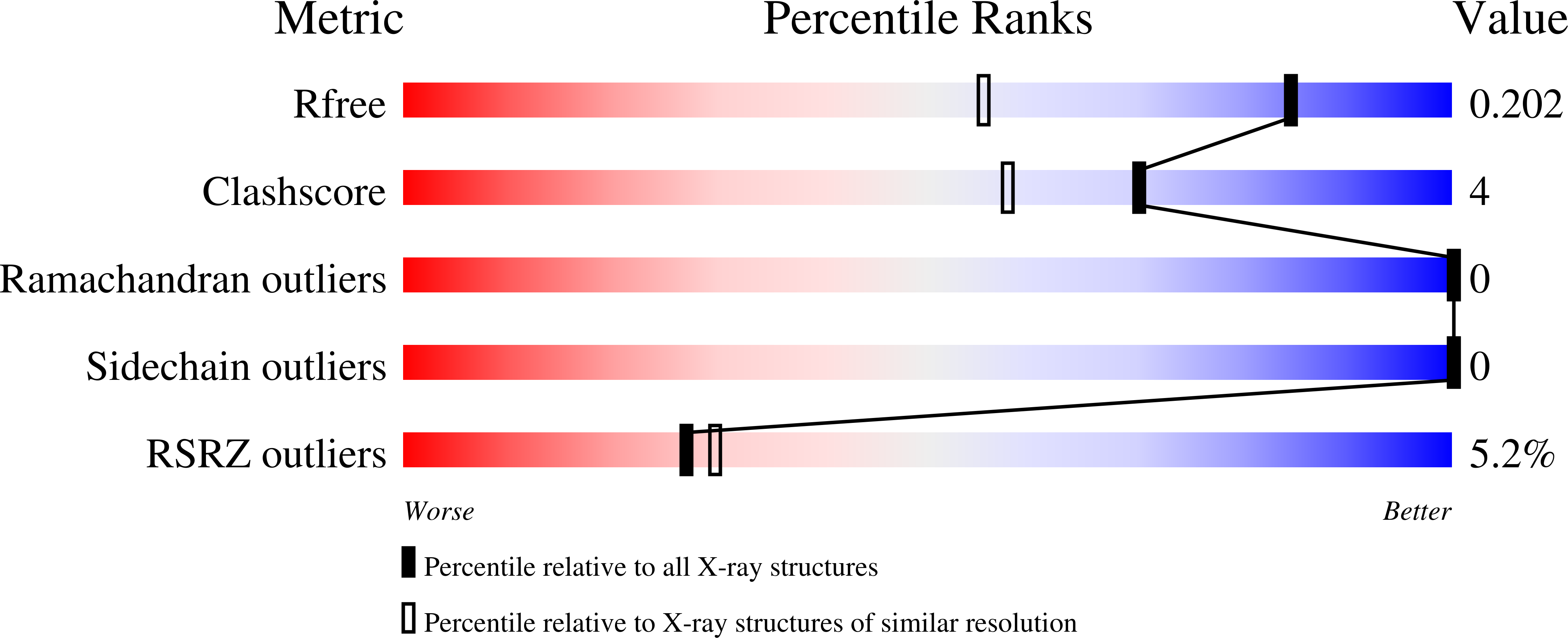
wwPDB Validation   3D Report Full Report


Ligand Structure Quality Assessment 


This is version 1.3 of the entry. See complete history


Literature

The Macro Domain is an Adp-Ribose Binding Module.

Karras, G.I.Kustatscher, G.Buhecha, H.R.Allen, M.D.Pugieux, C.Sait, F.Bycroft, M.Ladurner, A.G.

(2005) EMBO J 24: 1911

  • DOI: https://doi.org/10.1038/sj.emboj.7600664
  • Primary Citation of Related Structures:  
    2BFQ, 2BFR

  • PubMed Abstract: 

    The ADP-ribosylation of proteins is an important post-translational modification that occurs in a variety of biological processes, including DNA repair, transcription, chromatin biology and long-term memory formation. Yet no protein modules are known that specifically recognize the ADP-ribose nucleotide. We provide biochemical and structural evidence that macro domains are high-affinity ADP-ribose binding modules. Our structural analysis reveals a conserved ligand binding pocket among the macro domain fold. Consistently, distinct human macro domains retain their ability to bind ADP-ribose. In addition, some macro domain proteins also recognize poly-ADP-ribose as a ligand. Our data suggest an important role for proteins containing macro domains in the biology of ADP-ribose.


  • Organizational Affiliation

    Gene Expression Programme and Structural & Computational Biology Programme, European Molecular Biology Laboratory, Heidelberg, Germany.


Macromolecules
Find similar proteins by:  (by identity cutoff)  |  3D Structure
Entity ID: 1
MoleculeChains Sequence LengthOrganismDetailsImage
HYPOTHETICAL PROTEIN AF1521192Archaeoglobus fulgidusMutation(s): 0 
UniProt
Find proteins for O28751 (Archaeoglobus fulgidus (strain ATCC 49558 / DSM 4304 / JCM 9628 / NBRC 100126 / VC-16))
Explore O28751 
Go to UniProtKB:  O28751
Entity Groups  
Sequence Clusters30% Identity50% Identity70% Identity90% Identity95% Identity100% Identity
UniProt GroupO28751
Sequence Annotations
Expand
  • Reference Sequence
Small Molecules
Ligands 1 Unique
IDChains Name / Formula / InChI Key2D Diagram3D Interactions
AR6
Query on AR6

Download Ideal Coordinates CCD File 
B [auth A][(2R,3S,4R,5R)-5-(6-AMINOPURIN-9-YL)-3,4-DIHYDROXY-OXOLAN-2-YL]METHYL [HYDROXY-[[(2R,3S,4R,5S)-3,4,5-TRIHYDROXYOXOLAN-2-YL]METHOXY]PHOSPHORYL] HYDROGEN PHOSPHATE
C15 H23 N5 O14 P2
SRNWOUGRCWSEMX-ZQSHOCFMSA-N
Modified Residues  1 Unique
IDChains TypeFormula2D DiagramParent
FME
Query on FME
A
L-PEPTIDE LINKINGC6 H11 N O3 SMET
Binding Affinity Annotations 
IDSourceBinding Affinity
APR PDBBind:  2BFQ Kd: 130 (nM) from 1 assay(s)
Experimental Data & Validation

Experimental Data

  • Method: X-RAY DIFFRACTION
  • Resolution: 1.50 Å
  • R-Value Free: 0.233 
  • R-Value Work: 0.210 
  • R-Value Observed: 0.210 
  • Space Group: P 61
Unit Cell:
Length ( Å )Angle ( ˚ )
a = 88.116α = 90
b = 88.116β = 90
c = 60.28γ = 120
Software Package:
Software NamePurpose
CNSrefinement
MOSFLMdata reduction
SCALAdata scaling
MOLREPphasing

Structure Validation

View Full Validation Report


Ligand Structure Quality Assessment 


Entry History 

Deposition Data

Revision History  (Full details and data files)

  • Version 1.0: 2005-01-18
    Type: Initial release
  • Version 1.1: 2012-02-22
    Changes: Atomic model, Data collection, Derived calculations, Non-polymer description, Other, Refinement description, Structure summary, Version format compliance
  • Version 1.2: 2018-01-24
    Changes: Source and taxonomy
  • Version 1.3: 2023-12-13
    Changes: Data collection, Database references, Derived calculations, Other, Refinement description, Structure summary