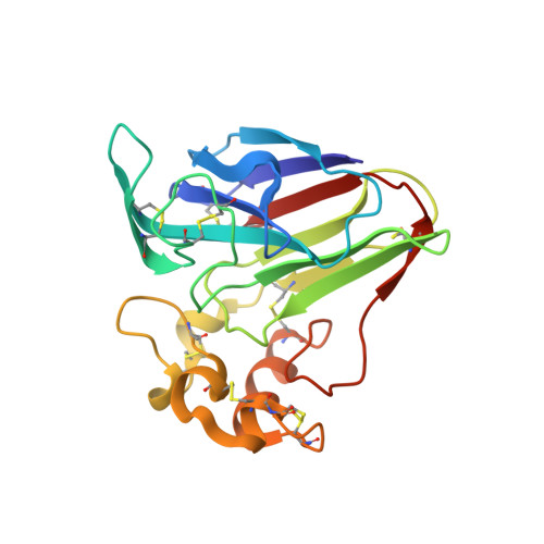
Improving Radiation-Damage Substructures for Rip.
Nanao, M.H., Sheldrick, G.M., Ravelli, R.B.(2005) Acta Crystallogr D Biol Crystallogr 61: 1227
- PubMed: 16131756
- DOI: https://doi.org/10.1107/S0907444905019360
- Primary Citation of Related Structures:
2BLO, 2BLP, 2BLQ, 2BLR, 2BLU, 2BLV, 2BLW, 2BLX, 2BLY, 2BLZ, 2BN1, 2BN3 - PubMed Abstract:
Specific radiation damage can be used to solve macromolecular structures using the radiation-damage-induced phasing (RIP) method. The method has been investigated for six disulfide-containing test structures (elastase, insulin, lysozyme, ribonuclease A, trypsin and thaumatin) using data sets that were collected on a third-generation synchrotron undulator beamline with a highly attenuated beam. Each crystal was exposed to the unattenuated X-ray beam between the collection of a 'before' and an 'after' data set. The X-ray 'burn'-induced intensity differences ranged from 5 to 15%, depending on the protein investigated. X-ray-susceptible substructures were determined using the integrated direct and Patterson methods in SHELXD. The best substructures were found by downscaling the 'after' data set in SHELXC by a scale factor K, with optimal values ranging from 0.96 to 0.99. The initial substructures were improved through iteration with SHELXE by the addition of negatively occupied sites as well as a large number of relatively weak sites. The final substructures ranged from 40 to more than 300 sites, with strongest peaks as high as 57sigma. All structures except one could be solved: it was not possible to find the initial substructure for ribonuclease A, however, SHELXE iteration starting with the known five most susceptible sites gave excellent maps. Downscaling proved to be necessary for the solution of elastase, lysozyme and thaumatin and reduced the number of SHELXE iterations in the other cases. The combination of downscaling and substructure iteration provides important benefits for the phasing of macromolecular structures using radiation damage.
Organizational Affiliation:
EMBL, 6 Rue Jules Horowitz, 38042 Grenoble, France.


