2BY0

Is radiation damage dependent on the dose-rate used during macromolecular crystallography data collection


Experimental Data Snapshot

  • Method: X-RAY DIFFRACTION
  • Resolution: 1.55 Å
  • R-Value Free: 
    0.175 (Depositor), 0.170 (DCC) 
  • R-Value Work: 
    0.146 (Depositor), 0.150 (DCC) 
  • R-Value Observed: 
    0.146 (Depositor) 

wwPDB Validation   3D Report Full Report


Ligand Structure Quality Assessment 

Created with Raphaël 2.3.0Worse 01 BetterLigand structure goodness of fit to experimental dataBest fitted GLCClick on this verticalbar to view details

This is version 2.0 of the entry. See complete history


Literature

Is Radiation Damage Dependent on the Dose-Rate Used During Macromolecular Crystallography Data Collection?

Leiros, H.-K.S.Timmins, J.Ravelli, R.B.G.Mcsweeney, S.M.

(2006) Acta Crystallogr D Biol Crystallogr 62: 125

  • DOI: https://doi.org/10.1107/S0907444905033627
  • Primary Citation of Related Structures:  
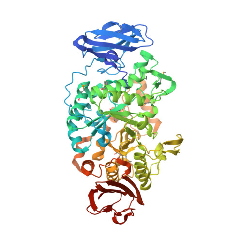
    2BXY, 2BXZ, 2BY0, 2BY1, 2BY2, 2BY3, 2BY5, 2BY6, 2BY7, 2BY8, 2BY9, 2BYA

  • PubMed Abstract: 

    This paper focuses on the radiation-damage effects when applying the same total X-ray dose to protein crystals at different dose rates. These experiments have been performed on both a selenomethionated protein and on bovine trypsin using dose rates that span nearly two orders of magnitude. The results show no clear dose-rate effect on the global indicators of radiation damage, but a small measurable dose-rate effect could be found when studying specific radiation damage. It is hypothesized that this observed dose-rate effect relates to differences in the steady-state free-radical concentration.


  • Organizational Affiliation

    Macromolecular Crystallography Group, European Synchrotron Radiation Facility (ESRF), 6 Rue Jules Horowitz, 38043 Grenoble, France.


Macromolecules
Find similar proteins by:  (by identity cutoff)  |  3D Structure
Entity ID: 1
MoleculeChains Sequence LengthOrganismDetailsImage
MALTOOLIGOSYLTREHALOSE TREHALOHYDROLASE602Deinococcus radiodurans R1 = ATCC 13939 = DSM 20539Mutation(s): 0 
EC: 3.2.1.1 (PDB Primary Data), 3.2.1.141 (UniProt)
UniProt
Find proteins for Q9RX51 (Deinococcus radiodurans (strain ATCC 13939 / DSM 20539 / JCM 16871 / CCUG 27074 / LMG 4051 / NBRC 15346 / NCIMB 9279 / VKM B-1422 / R1))
Explore Q9RX51 
Go to UniProtKB:  Q9RX51
Entity Groups  
Sequence Clusters30% Identity50% Identity70% Identity90% Identity95% Identity100% Identity
UniProt GroupQ9RX51
Sequence Annotations
Expand
  • Reference Sequence
Oligosaccharides

Help

Entity ID: 2
MoleculeChains Length2D Diagram Glycosylation3D Interactions
alpha-D-glucopyranose-(1-1)-alpha-D-glucopyranose
B, C
2N/A
Glycosylation Resources
GlyTouCan:  G92130SN
GlyCosmos:  G92130SN
Small Molecules
Biologically Interesting Molecules (External Reference) 1 Unique
Experimental Data & Validation

Experimental Data

  • Method: X-RAY DIFFRACTION
  • Resolution: 1.55 Å
  • R-Value Free:  0.175 (Depositor), 0.170 (DCC) 
  • R-Value Work:  0.146 (Depositor), 0.150 (DCC) 
  • R-Value Observed: 0.146 (Depositor) 
Space Group: P 21 21 21
Unit Cell:
Length ( Å )Angle ( ˚ )
a = 59.511α = 90
b = 66.646β = 90
c = 153.1γ = 90
Software Package:
Software NamePurpose
REFMACrefinement
MOSFLMdata reduction
SCALAdata scaling

Structure Validation

View Full Validation Report


Ligand Structure Quality Assessment 

Created with Raphaël 2.3.0Worse 01 BetterLigand structure goodness of fit to experimental dataBest fitted GLCClick on this verticalbar to view details

Entry History 

Deposition Data

Revision History  (Full details and data files)

  • Version 1.0: 2006-02-06
    Type: Initial release
  • Version 1.1: 2011-05-08
    Changes: Version format compliance
  • Version 1.2: 2011-07-13
    Changes: Version format compliance
  • Version 2.0: 2020-07-29
    Type: Remediation
    Reason: Carbohydrate remediation
    Changes: Advisory, Atomic model, Data collection, Derived calculations, Non-polymer description, Other, Structure summary