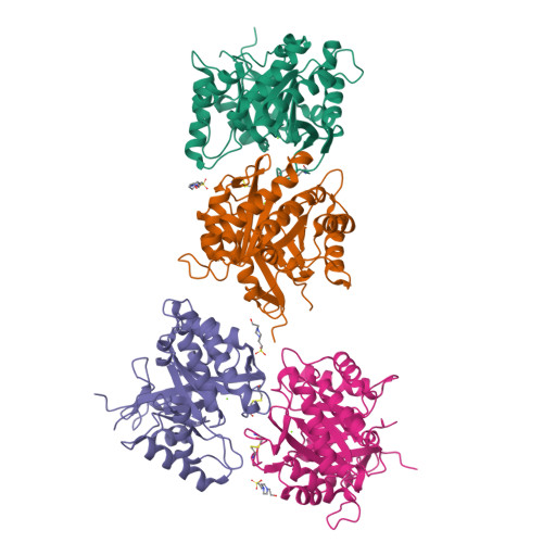
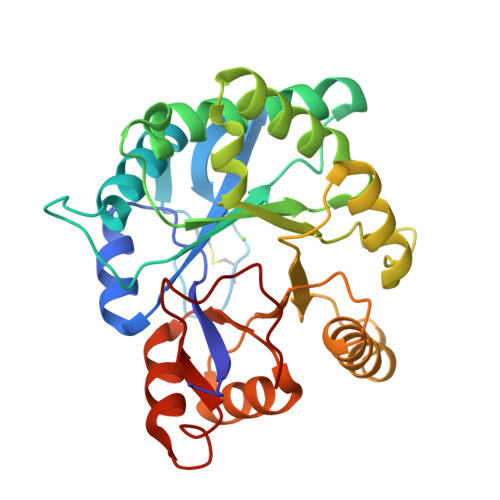
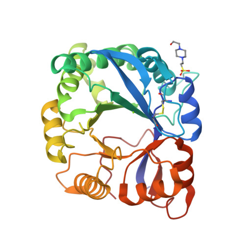
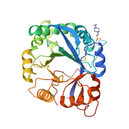
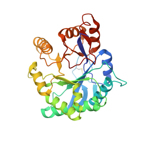
2F9R

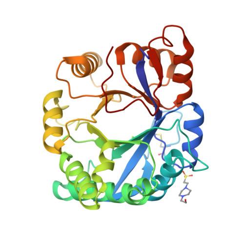
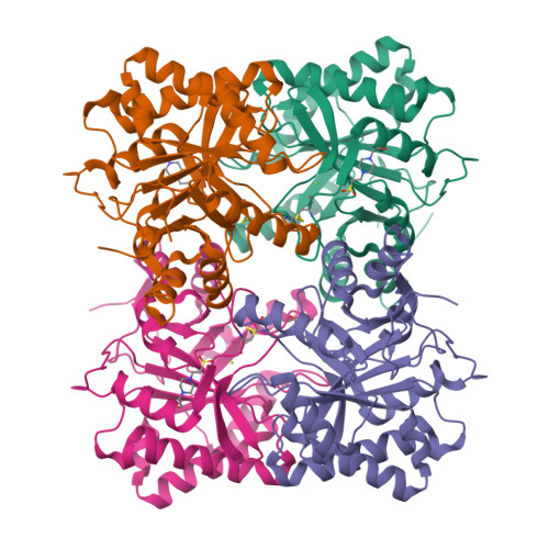
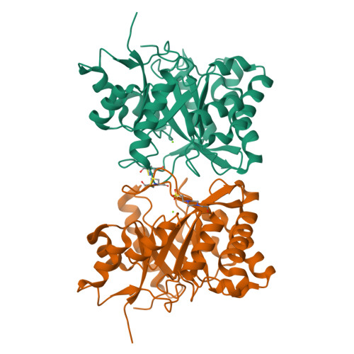
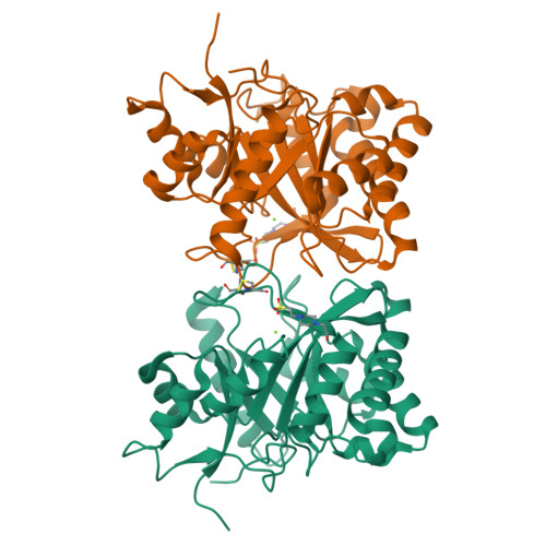
Crystal structure of the inactive state of the Smase I, a sphingomyelinase D from Loxosceles laeta venom


Experimental Data Snapshot

  • Method: X-RAY DIFFRACTION
  • Resolution: 1.85 Å
  • R-Value Free: 0.234 
  • R-Value Work: 0.187 
  • R-Value Observed: 0.190 

Starting Model: experimental
View more details

wwPDB Validation   3D Report Full Report


This is version 1.4 of the entry. See complete history


Literature

Structural basis for metal ion coordination and the catalytic mechanism of sphingomyelinases D.

Murakami, M.T.Gabdoulkhakov, A.Fernandes-Pedrosa, M.F.Tambourgi, D.V.Arni, R.K.

(2005) J Biol Chem 280: 13658-13664

  • DOI: https://doi.org/10.1074/jbc.M412437200
  • Primary Citation of Related Structures:  
    1XX1, 2F9R

  • PubMed Abstract: 

    Sphingomyelinases D (SMases D) from Loxosceles spider venom are the principal toxins responsible for the manifestation of dermonecrosis, intravascular hemolysis, and acute renal failure, which can result in death. These enzymes catalyze the hydrolysis of sphingomyelin, resulting in the formation of ceramide 1-phosphate and choline or the hydrolysis of lysophosphatidyl choline, generating the lipid mediator lysophosphatidic acid. This report represents the first crystal structure of a member of the sphingomyelinase D family from Loxosceles laeta (SMase I), which has been determined at 1.75-angstrom resolution using the "quick cryo-soaking" technique and phases obtained from a single iodine derivative and data collected from a conventional rotating anode x-ray source. SMase I folds as an (alpha/beta)8 barrel, the interfacial and catalytic sites encompass hydrophobic loops and a negatively charged surface. Substrate binding and/or the transition state are stabilized by a Mg2+ ion, which is coordinated by Glu32, Asp34, Asp91, and solvent molecules. In the proposed acid base catalytic mechanism, His12 and His47 play key roles and are supported by a network of hydrogen bonds between Asp34, Asp52, Trp230, Asp233, and Asn252.


  • Organizational Affiliation

    Department of Physics, Instituto de Biociências, Letras e Ciências Exatas/Universidade Estadual Paulista, São José do Rio Preto, SP 15054-000, Brazil.


Macromolecules
Find similar proteins by:  (by identity cutoff)  |  3D Structure
Entity ID: 1
MoleculeChains Sequence LengthOrganismDetailsImage
Sphingomyelinase D 1
A, B, C, D
285Loxosceles laetaMutation(s): 0 
EC: 3.1.4.41 (PDB Primary Data), 4.6.1 (UniProt)
UniProt
Find proteins for Q8I914 (Loxosceles laeta)
Explore Q8I914 
Go to UniProtKB:  Q8I914
Entity Groups  
Sequence Clusters30% Identity50% Identity70% Identity90% Identity95% Identity100% Identity
UniProt GroupQ8I914
Sequence Annotations
Expand
  • Reference Sequence
Experimental Data & Validation

Experimental Data

  • Method: X-RAY DIFFRACTION
  • Resolution: 1.85 Å
  • R-Value Free: 0.234 
  • R-Value Work: 0.187 
  • R-Value Observed: 0.190 
  • Space Group: P 65
Unit Cell:
Length ( Å )Angle ( ˚ )
a = 140.587α = 90
b = 140.587β = 90
c = 113.608γ = 120
Software Package:
Software NamePurpose
REFMACrefinement
DENZOdata reduction
SCALEPACKdata scaling
AMoREphasing

Structure Validation

View Full Validation Report


Entry History 

Deposition Data

Revision History  (Full details and data files)

  • Version 1.0: 2006-06-27
    Type: Initial release
  • Version 1.1: 2008-05-01
    Changes: Version format compliance
  • Version 1.2: 2011-07-13
    Changes: Derived calculations, Version format compliance
  • Version 1.3: 2023-08-30
    Changes: Data collection, Database references, Derived calculations, Refinement description
  • Version 1.4: 2024-10-30
    Changes: Structure summary