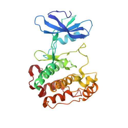
2FH9

Structure and dimerization of the kinase domain from yeast Snf1


Experimental Data Snapshot

  • Method: X-RAY DIFFRACTION
  • Resolution: 2.80 Å
  • R-Value Free: 0.260 
  • R-Value Work: 0.215 
  • R-Value Observed: 0.336 

wwPDB Validation   3D Report Full Report


This is version 1.5 of the entry. See complete history


Literature

Structure and Dimerization of the Kinase Domain from Yeast Snf1, a Member of the Snf1/AMPK Protein Family

Nayak, V.Zhao, K.Wyce, A.Schwartz, M.F.Lo, W.S.Berger, S.L.Marmorstein, R.

(2006) Structure 14: 477-485

  • DOI: https://doi.org/10.1016/j.str.2005.12.008
  • Primary Citation of Related Structures:  
    2FH9

  • PubMed Abstract: 

    The Snf1/AMPK kinases are intracellular energy sensors, and the AMPK pathway has been implicated in a variety of metabolic human disorders. Here we report the crystal structure of the kinase domain from yeast Snf1, revealing a bilobe kinase fold with greatest homology to cyclin-dependant kinase-2. Unexpectedly, the crystal structure also reveals a novel homodimer that we show also forms in solution, as demonstrated by equilibrium sedimentation, and in yeast cells, as shown by coimmunoprecipitation of differentially tagged intact Snf1. A mapping of sequence conservation suggests that dimer formation is a conserved feature of the Snf1/AMPK kinases. The conformation of the conserved alphaC helix, and the burial of the activation segment and substrate binding site within the dimer, suggests that it represents an inactive form of the kinase. Taken together, these studies suggest another layer of kinase regulation within the Snf1/AMPK family, and an avenue for development of AMPK-specific activating compounds.


  • Organizational Affiliation

    The Wistar Institute, University of Pennsylvania, Philadelphia 19104, USA.


Macromolecules
Find similar proteins by:  (by identity cutoff)  |  3D Structure
Entity ID: 1
MoleculeChains Sequence LengthOrganismDetailsImage
Snf1 kinase274Saccharomyces cerevisiaeMutation(s): 0 
Gene Names: SNF1CAT1CCR1GLC2PAS14
EC: 2.7.1 (PDB Primary Data), 2.7.11.1 (UniProt)
UniProt
Find proteins for P06782 (Saccharomyces cerevisiae (strain ATCC 204508 / S288c))
Explore P06782 
Go to UniProtKB:  P06782
Entity Groups  
Sequence Clusters30% Identity50% Identity70% Identity90% Identity95% Identity100% Identity
UniProt GroupP06782
Sequence Annotations
Expand
  • Reference Sequence
Experimental Data & Validation

Experimental Data

  • Method: X-RAY DIFFRACTION
  • Resolution: 2.80 Å
  • R-Value Free: 0.260 
  • R-Value Work: 0.215 
  • R-Value Observed: 0.336 
  • Space Group: P 4 21 2
Unit Cell:
Length ( Å )Angle ( ˚ )
a = 108.677α = 90
b = 108.677β = 90
c = 61.62γ = 90
Software Package:
Software NamePurpose
DENZOdata reduction
SCALEPACKdata scaling
SOLVEphasing
RESOLVEphasing
CNSrefinement
PDB_EXTRACTdata extraction

Structure Validation

View Full Validation Report


Entry History 

Deposition Data

  • Released Date: 2006-03-28 
  • Deposition Author(s): Nayak, V.

Revision History  (Full details and data files)

  • Version 1.0: 2006-03-28
    Type: Initial release
  • Version 1.1: 2008-05-01
    Changes: Version format compliance
  • Version 1.2: 2011-07-13
    Changes: Derived calculations, Version format compliance
  • Version 1.3: 2011-11-16
    Changes: Atomic model
  • Version 1.4: 2017-10-18
    Changes: Refinement description
  • Version 1.5: 2024-02-14
    Changes: Data collection, Database references