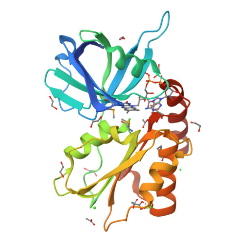
Crystal structure of siderophore-interacting protein (ZP_00813641.1) from Shewanella putrefaciens CN-32 at 2.20 A resolution
Joint Center for Structural Genomics (JCSG)To be published.
Experimental Data Snapshot
Entity ID: 1 | |||||
|---|---|---|---|---|---|
| Molecule | Chains | Sequence Length | Organism | Details | Image |
| Siderophore-interacting protein | 252 | Shewanella putrefaciens CN-32 | Mutation(s): 6 Gene Names: ZP_00813641.1 |  | |
UniProt | |||||
Find proteins for A4Y1H9 (Shewanella putrefaciens (strain CN-32 / ATCC BAA-453)) Explore A4Y1H9 Go to UniProtKB: A4Y1H9 | |||||
Entity Groups | |||||
| Sequence Clusters | 30% Identity50% Identity70% Identity90% Identity95% Identity100% Identity | ||||
| UniProt Group | A4Y1H9 | ||||
Sequence AnnotationsExpand | |||||
| |||||
| Ligands 5 Unique | |||||
|---|---|---|---|---|---|
| ID | Chains | Name / Formula / InChI Key | 2D Diagram | 3D Interactions | |
| FAD Query on FAD | E [auth A] | FLAVIN-ADENINE DINUCLEOTIDE C27 H33 N9 O15 P2 VWWQXMAJTJZDQX-UYBVJOGSSA-N |  | ||
| EDO Query on EDO | F [auth A] G [auth A] H [auth A] I [auth A] J [auth A] | 1,2-ETHANEDIOL C2 H6 O2 LYCAIKOWRPUZTN-UHFFFAOYSA-N |  | ||
| ACY Query on ACY | N [auth A] O [auth A] P [auth A] Q [auth A] R [auth A] | ACETIC ACID C2 H4 O2 QTBSBXVTEAMEQO-UHFFFAOYSA-N |  | ||
| CA Query on CA | B [auth A], C [auth A] | CALCIUM ION Ca BHPQYMZQTOCNFJ-UHFFFAOYSA-N |  | ||
| CL Query on CL | D [auth A] | CHLORIDE ION Cl VEXZGXHMUGYJMC-UHFFFAOYSA-M |  | ||
| Modified Residues 1 Unique | |||||
|---|---|---|---|---|---|
| ID | Chains | Type | Formula | 2D Diagram | Parent |
| MSE Query on MSE | A | L-PEPTIDE LINKING | C5 H11 N O2 Se |  | MET |
| Length ( Å ) | Angle ( ˚ ) |
|---|---|
| a = 100.54 | α = 90 |
| b = 100.54 | β = 90 |
| c = 67.51 | γ = 90 |
| Software Name | Purpose |
|---|---|
| REFMAC | refinement |
| XSCALE | data scaling |
| PDB_EXTRACT | data extraction |
| XDS | data reduction |
| autoSHARP | phasing |
RCSB PDB is hosted by

RCSB PDB is a member of the