2GUY

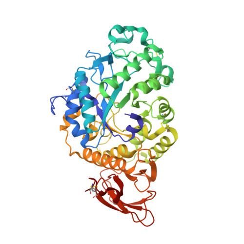
Orthorhombic crystal structure (space group P21212) of Aspergillus niger alpha-amylase at 1.6 A resolution


Experimental Data Snapshot

  • Method: X-RAY DIFFRACTION
  • Resolution: 1.59 Å
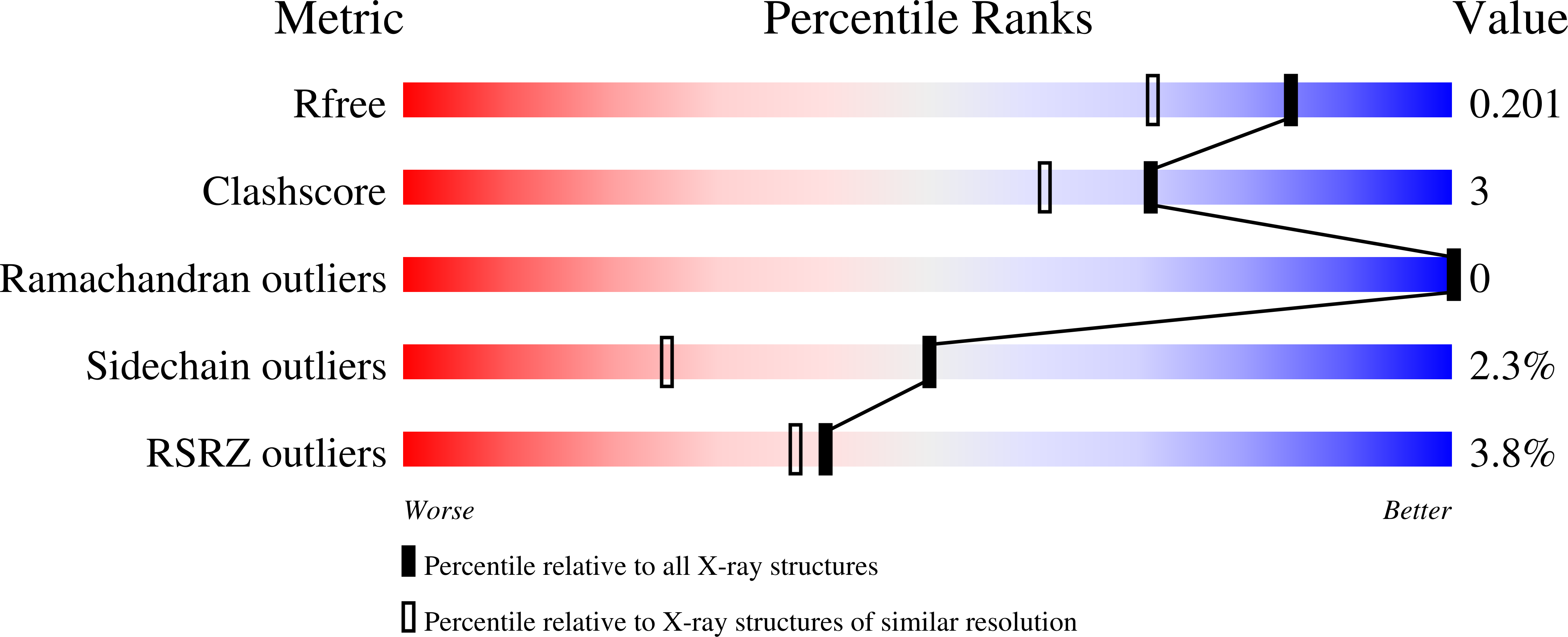
  • R-Value Free: 0.196 
  • R-Value Work: 0.164 
  • R-Value Observed: 0.165 

Starting Model: experimental
View more details

wwPDB Validation   3D Report Full Report


This is version 2.2 of the entry. See complete history


Literature

Monoclinic crystal form of Aspergillus niger alpha-amylase in complex with maltose at 1.8 angstroms resolution.

Vujicic-Zagar, A.Dijkstra, B.W.

(2006) Acta Crystallogr Sect F Struct Biol Cryst Commun 62: 716-721

  • DOI: https://doi.org/10.1107/S1744309106024729
  • Primary Citation of Related Structures:  
    2GUY, 2GVY

  • PubMed Abstract: 

    Aspergillus niger alpha-amylase catalyses the hydrolysis of alpha-1,4-glucosidic bonds in starch. It shows 100% sequence identity to the A. oryzae homologue (also called TAKA-amylase), three crystal structures of which have been published to date. Two of them belong to the orthorhombic space group P2(1)2(1)2(1) with one molecule per asymmetric unit and one belongs to the monoclinic space group P2(1) with three molecules per asymmetric unit. Here, the purification, crystallization and structure determination of A. niger alpha-amylase crystallized in the monoclinic space group P2(1) with two molecules per asymmetric unit in complex with maltose at 1.8 angstroms resolution is reported. Furthermore, a novel 1.6 angstroms resolution orthorhombic crystal form (space group P2(1)2(1)2) of the native enzyme is presented. Four maltose molecules are observed in the maltose-alpha-amylase complex. Three of these occupy active-site subsites -2 and -1, +1 and +2 and the hitherto unobserved subsites +4 (Asp233, Gly234) and +5 (Asp235). The fourth maltose molecule binds at the distant binding sites d1 (Tyr382) and d2 (Trp385), also previously unobserved. Furthermore, it is shown that the active-site groove permits different binding modes of sugar units at subsites +1 and +2. This flexibility of the active-site cleft close to the catalytic centre might be needed for a productive binding of substrate chains and/or release of products.


  • Organizational Affiliation

    Laboratory of Biophysical Chemistry, University of Groningen, Nijenborgh 4, 9747AG Groningen, The Netherlands.


Macromolecules
Find similar proteins by:  (by identity cutoff)  |  3D Structure
Entity ID: 1
MoleculeChains Sequence LengthOrganismDetailsImage
Alpha-amylase A478Aspergillus oryzaeMutation(s): 0 
EC: 3.2.1.1
UniProt
Find proteins for P0C1B3 (Aspergillus oryzae (strain ATCC 42149 / RIB 40))
Explore P0C1B3 
Go to UniProtKB:  P0C1B3
Entity Groups  
Sequence Clusters30% Identity50% Identity70% Identity90% Identity95% Identity100% Identity
UniProt GroupP0C1B3
Glycosylation
Glycosylation Sites: 1
Sequence Annotations
Expand
  • Reference Sequence
Oligosaccharides

Help

Entity ID: 2
MoleculeChains Length2D Diagram Glycosylation3D Interactions
beta-D-mannopyranose-(1-4)-2-acetamido-2-deoxy-beta-D-glucopyranose-(1-4)-2-acetamido-2-deoxy-beta-D-glucopyranose
B
3N-Glycosylation
Glycosylation Resources
GlyTouCan:  G15407YE
GlyCosmos:  G15407YE
GlyGen:  G15407YE
Small Molecules
Ligands 1 Unique
IDChains Name / Formula / InChI Key2D Diagram3D Interactions
CA
Query on CA

Download Ideal Coordinates CCD File 
C [auth A]CALCIUM ION
Ca
BHPQYMZQTOCNFJ-UHFFFAOYSA-N
Experimental Data & Validation

Experimental Data

  • Method: X-RAY DIFFRACTION
  • Resolution: 1.59 Å
  • R-Value Free: 0.196 
  • R-Value Work: 0.164 
  • R-Value Observed: 0.165 
  • Space Group: P 21 21 2
Unit Cell:
Length ( Å )Angle ( ˚ )
a = 102.799α = 90
b = 63.228β = 90
c = 74.456γ = 90
Software Package:
Software NamePurpose
REFMACrefinement
ProDCdata collection
SCALEPACKdata scaling
PHASERphasing

Structure Validation

View Full Validation Report


Entry History 

Deposition Data

Revision History  (Full details and data files)

  • Version 1.0: 2006-08-15
    Type: Initial release
  • Version 1.1: 2008-05-01
    Changes: Version format compliance
  • Version 1.2: 2011-07-13
    Changes: Advisory, Version format compliance
  • Version 1.3: 2017-10-18
    Changes: Refinement description
  • Version 2.0: 2020-07-29
    Type: Remediation
    Reason: Carbohydrate remediation
    Changes: Atomic model, Data collection, Derived calculations, Structure summary
  • Version 2.1: 2023-08-30
    Changes: Data collection, Database references, Refinement description, Structure summary
  • Version 2.2: 2024-11-06
    Changes: Structure summary