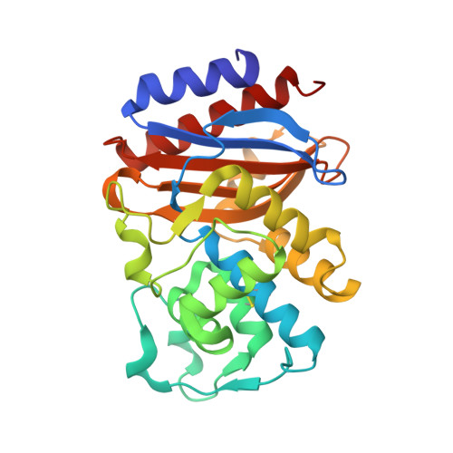
Effect of the inhibitor-resistant M69V substitution on the structures and populations of trans-enamine beta-lactamase intermediates.
Totir, M.A., Padayatti, P.S., Helfand, M.S., Carey, M.P., Bonomo, R.A., Carey, P.R., van den Akker, F.(2006) Biochemistry 45: 11895-11904
- PubMed: 17002290
- DOI: https://doi.org/10.1021/bi060990m
- Primary Citation of Related Structures:
2H0T, 2H0Y, 2H10 - PubMed Abstract:
The objective of this study was to determine the molecular factors that lead to beta-lactamase inhibitor resistance for the M69V variant in SHV-1 beta-lactamase. With mechanism-based inhibitors, the beta-lactamase forms an acyl-enzyme intermediate that consists of a trans-enamine derivative in the active site. This study focuses on these intermediates by introducing the E166A mutation that greatly retards deacylation. Thus, by comparing the properties of the E166A and M69V/E166A forms, we can explore the consequences of the resistance mutation at the level of the enamine acyl-enzyme forms. The reactions between the beta-lactamase and the inhibitors tazobactam, sulbactam, and clavulanic acid are followed in single crystals of the enzymes by using a Raman microscope. The resulting Raman difference spectroscopic data provide detailed information about conformational events involving the enamine species as well as an estimate of their populations. The Raman difference spectra for each of the inhibitors in the E166A and M69V/E166A variants are very similar. In particular, detailed analysis of the main enamine Raman vibration near 1595 cm(-1) reveals that the structure and flexibility of the enamine fragments are essentially identical for each of the three inhibitors in E166A and in the M69V/E166A double mutant. This finding is in accord with the X-ray-derived structures, presented herein at 1.6-1.75 A resolution, of the trans-enamine intermediates formed by the three inhibitors in M69V/E166A. However, a comparison of Raman results for M69V/E166A and E166A shows that the M69V mutation results in a 40%, 25%, and negligible reductions in the enamine population when the beta-lactamase crystals are soaked in 5 mM tazobactam, clavulanic acid, and sulbactam solutions, respectively. The levels of enamine from tazobactam and clavulanic acid can be increased by increasing the concentrations of inhibitor in the mother liquor. Thus, the sensitivity of population levels to the inhibitor concentration in the mother liquor focuses attention on the properties of the encounter complex preceding acylation. It is proposed that for small ligands, such as tazobactam, sulbactam, and clavulanic acid, the positioning of the lactam ring in the active site in the correct orientation for acylation is only one of a number of poorly defined conformations. For tazobactam and clavulanic acid, the correctly oriented encounter complex is even less likely in the M69V variant, leading to a reduction in the level of inhibition of the enzyme via formation of the acyl-enzyme intermediate and the onset of resistance. Analysis of the X-ray structures of the three intermediates in M69V/E166A demonstrates that, compared to the structures for the E166A form, the oxyanion hole becomes smaller, providing one explanation for why acylation may be less efficient following the M69V substitution.
Organizational Affiliation:
Department of Biochemistry, Case Western Reserve University, Cleveland, Ohio 44106, USA.


