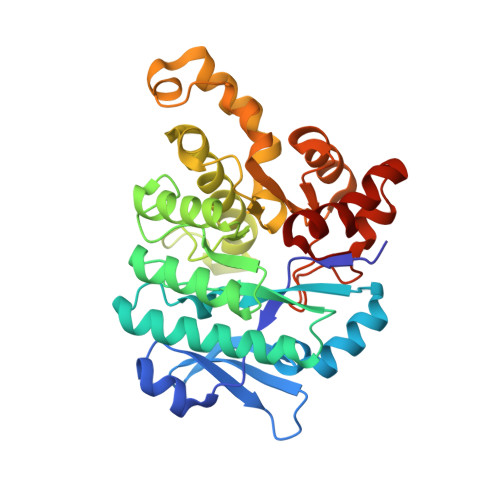
Crystal Structure of alpha-Amino-beta-carboxymuconate-epsilon-semialdehyde Decarboxylase: Insight into the Active Site and Catalytic Mechanism of a Novel Decarboxylation Reaction.
Martynowski, D., Eyobo, Y., Li, T., Yang, K., Liu, A., Zhang, H.(2006) Biochemistry 45: 10412-10421
- PubMed: 16939194
- DOI: https://doi.org/10.1021/bi060903q
- Primary Citation of Related Structures:
2HBV, 2HBX - PubMed Abstract:
Alpha-amino-beta-carboxymuconate-epsilon-semialdehyde decarboxylase (ACMSD) is a widespread enzyme found in many bacterial species and all currently sequenced eukaryotic organisms. It occupies a key position at the branching point of two metabolic pathways: the tryptophan to quinolinate pathway and the bacterial 2-nitrobenzoic acid degradation pathway. The activity of ACMSD determines whether the metabolites in both pathways are converted to quinolinic acid for NAD biosynthesis or to acetyl-CoA for the citric acid cycle. Here we report the first high-resolution crystal structure of ACMSD from Pseudomonas fluorescens which validates our previous predictions that this enzyme is a member of the metal-dependent amidohydrolase superfamily of the (beta/alpha)(8) TIM barrel fold. The structure of the enzyme in its native form, determined at 1.65 A resolution, reveals the precise spatial arrangement of the active site metal center and identifies a potential substrate-binding pocket. The identity of the native active site metal was determined to be Zn. Also determined was the structure of the enzyme complexed with cobalt at 2.50 A resolution. The hydrogen bonding network around the metal center suggests that Arg51 and His228 may play important roles in catalysis. The metal center configuration of PfACMSD is very similar to that of Zn-dependent adenosine deaminase and Fe-dependent cytosine deaminase, suggesting that ACMSD may share certain similarities in its catalytic mechanism with these enzymes. These data enable us to propose possible catalytic mechanisms for ACMSD which appear to be unprecedented among all currently characterized decarboxylases.
Organizational Affiliation:
Department of Biochemistry, University of Texas Southwestern Medical Center, Dallas, Texas 75390-8816, USA.


