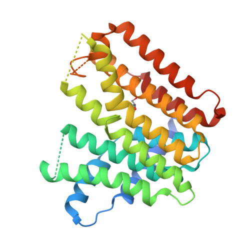
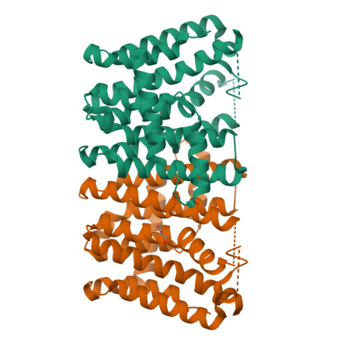
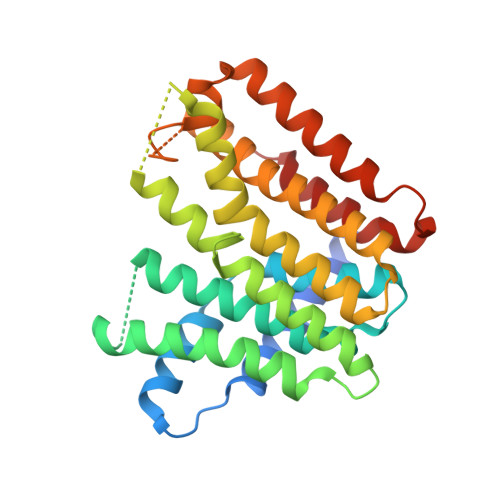
2J1O

Geranylgeranyl diphosphate synthase from Sinapis alba


Experimental Data Snapshot

  • Method: X-RAY DIFFRACTION
  • Resolution: 2.00 Å
  • R-Value Free: 0.230 
  • R-Value Work: 0.195 
  • R-Value Observed: 0.196 

wwPDB Validation   3D Report Full Report


This is version 1.2 of the entry. See complete history


Literature

Structure and Reaction Geometry of Geranylgeranyl Diphosphate Synthase from Sinapis Alba.

Kloer, D.P.Welsch, R.Beyer, P.Schulz, G.E.

(2006) Biochemistry 45: 15197

  • DOI: https://doi.org/10.1021/bi061572k
  • Primary Citation of Related Structures:  
    2J1O, 2J1P

  • PubMed Abstract: 

    The crystal structure of the geranylgeranyl diphosphate synthase from Sinapis alba (mustard) has been solved in two crystal forms at 1.8 and 2.0 A resolutions. In one of these forms, the dimeric enzyme binds one molecule of the final product geranylgeranyl diphosphate in one subunit. The chainfold of the enzyme corresponds to that of other members of the farnesyl diphosphate synthase family. Whereas the binding modes of the two substrates dimethylallyl diphosphate and isopentenyl diphosphate at the allyl and isopentenyl sites, respectively, have been established with other members of the family, the complex structure presented reveals for the first time the binding mode of a reaction product at the isopentenyl site. The binding geometry of substrates and product in conjunction with the protein environment and the established chemistry of the reaction provide a clear picture of the reaction steps and atom displacements. Moreover, a comparison with a ligated homologous structure outlined an appreciable induced fit: helix alpha8 and its environment undergo a large conformational change when either the substrate dimethylallyl diphosphate or an analogue is bound to the allyl site; only a minor conformational change occurs when the other substrate isopentenyl diphosphate or the product is bound to the isopentenyl site.


  • Organizational Affiliation

    Institut für Organische Chemie und Biochemie, Albert-Ludwigs-Universität, Albertstrasse 21, D-79104 Freiburg im Breisgau, Germany.


Macromolecules
Find similar proteins by:  (by identity cutoff)  |  3D Structure
Entity ID: 1
MoleculeChains Sequence LengthOrganismDetailsImage
GERANYLGERANYL PYROPHOSPHATE SYNTHETASE268Sinapis albaMutation(s): 0 
EC: 2.5.1.10 (UniProt), 2.5.1 (UniProt), 2.5.1.29 (UniProt), 2.5.1.1 (UniProt)
UniProt
Find proteins for Q43133 (Sinapis alba)
Explore Q43133 
Go to UniProtKB:  Q43133
Entity Groups  
Sequence Clusters30% Identity50% Identity70% Identity90% Identity95% Identity100% Identity
UniProt GroupQ43133
Sequence Annotations
Expand
  • Reference Sequence
Small Molecules
Ligands 1 Unique
IDChains Name / Formula / InChI Key2D Diagram3D Interactions
GOL
Query on GOL

Download Ideal Coordinates CCD File 
B [auth A]GLYCEROL
C3 H8 O3
PEDCQBHIVMGVHV-UHFFFAOYSA-N
Experimental Data & Validation

Experimental Data

  • Method: X-RAY DIFFRACTION
  • Resolution: 2.00 Å
  • R-Value Free: 0.230 
  • R-Value Work: 0.195 
  • R-Value Observed: 0.196 
  • Space Group: P 31 2 1
Unit Cell:
Length ( Å )Angle ( ˚ )
a = 71.81α = 90
b = 71.81β = 90
c = 127.85γ = 120
Software Package:
Software NamePurpose
REFMACrefinement
XDSdata reduction
XSCALEdata scaling

Structure Validation

View Full Validation Report


Entry History 

Deposition Data

Revision History  (Full details and data files)

  • Version 1.0: 2007-01-02
    Type: Initial release
  • Version 1.1: 2011-07-13
    Changes: Advisory, Version format compliance
  • Version 1.2: 2024-05-08
    Changes: Data collection, Database references, Other