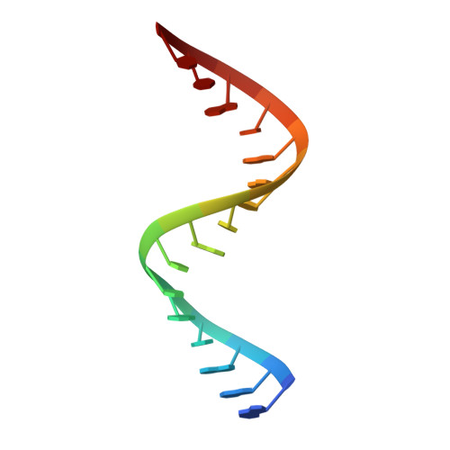
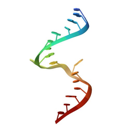
2OE6

2.4A X-ray crystal structure of unliganded RNA fragment GGGCGUCGCUAGUACC/CGGUACUAAAAGUCGCC containing the human ribosomal decoding A site: RNA construct with 5'-overhang


Experimental Data Snapshot

  • Method: X-RAY DIFFRACTION
  • Resolution: 2.40 Å
  • R-Value Free: 0.252 
  • R-Value Work: 0.209 
  • R-Value Observed: 0.212 

Starting Model: experimental
View more details

wwPDB Validation   3D Report Full Report


This is version 1.3 of the entry. See complete history


Literature

Apramycin recognition by the human ribosomal decoding site.

Hermann, T.Tereshko, V.Skripkin, E.Patel, D.J.

(2007) Blood Cells Mol Dis 38: 193-198

  • DOI: https://doi.org/10.1016/j.bcmd.2006.11.006
  • Primary Citation of Related Structures:  
    2OE5, 2OE6, 2OE8

  • PubMed Abstract: 

    Aminoglycoside antibiotics bind specifically to the bacterial ribosomal decoding-site RNA and thereby interfere with fidelity but not efficiency of translation. Apramycin stands out among aminoglycosides for its mechanism of action which is based on blocking translocation and its ability to bind also to the eukaryotic decoding site despite differences in key residues required for apramycin recognition by the bacterial target. To elucidate molecular recognition of the eukaryotic decoding site by apramycin we have determined the crystal structure of an oligoribonucleotide containing the human sequence free and in complex with the antibiotic at 1.5 A resolution. The drug binds in the deep groove of the RNA which forms a continuously stacked helix comprising non-canonical C.A and G.A base pairs and a bulged-out adenine. The binding mode of apramycin at the human decoding-site RNA is distinct from aminoglycoside recognition of the bacterial target, suggesting a molecular basis for the actions of apramycin in eukaryotes and bacteria.


  • Organizational Affiliation

    Department of Chemistry and Biochemistry, University of California-San Diego, La Jolla, CA 92093, USA. tch@ucsd.edu


Macromolecules

Find similar nucleic acids by:  Sequence   |   3D Structure  

Entity ID: 1
MoleculeChains LengthOrganismImage
RNA (5'-R(*GP*GP*GP*CP*GP*UP*CP*GP*CP*UP*AP*GP*UP*AP*CP*C)-3')16N/A
Sequence Annotations
Expand
  • Reference Sequence

Find similar nucleic acids by:  Sequence   |   3D Structure  

Entity ID: 2
MoleculeChains LengthOrganismImage
RNA (5'-R(*CP*GP*GP*UP*AP*CP*UP*AP*AP*AP*AP*GP*UP*CP*GP*CP*C)-3')17N/A
Sequence Annotations
Expand
  • Reference Sequence
Experimental Data & Validation

Experimental Data

  • Method: X-RAY DIFFRACTION
  • Resolution: 2.40 Å
  • R-Value Free: 0.252 
  • R-Value Work: 0.209 
  • R-Value Observed: 0.212 
  • Space Group: P 21 21 21
Unit Cell:
Length ( Å )Angle ( ˚ )
a = 28.157α = 90
b = 36.416β = 90
c = 87.325γ = 90
Software Package:
Software NamePurpose
REFMACrefinement
HKL-2000data collection
HKL-2000data reduction
HKL-2000data scaling
AMoREphasing

Structure Validation

View Full Validation Report


Entry History 

Deposition Data

Revision History  (Full details and data files)

  • Version 1.0: 2007-02-13
    Type: Initial release
  • Version 1.1: 2008-05-01
    Changes: Version format compliance
  • Version 1.2: 2011-07-13
    Changes: Advisory, Refinement description, Version format compliance
  • Version 1.3: 2023-08-30
    Changes: Data collection, Database references, Refinement description