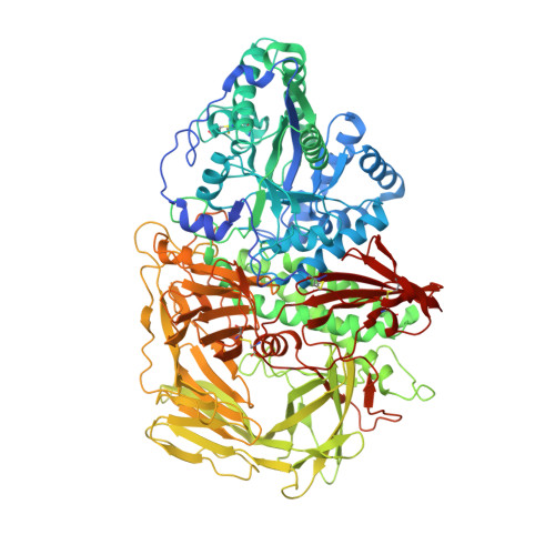
2OW7

Golgi alpha-mannosidase II complex with (1R,6S,7R,8S)-1-thioniabicyclo[4.3.0]nonan-7,8-diol chloride


Experimental Data Snapshot

  • Method: X-RAY DIFFRACTION
  • Resolution: 1.77 Å
  • R-Value Free: 0.203 
  • R-Value Work: 0.162 
  • R-Value Observed: 0.162 

Starting Model: experimental
View more details

wwPDB Validation   3D Report Full Report


Ligand Structure Quality Assessment 


This is version 1.4 of the entry. See complete history


Literature

Binding of sulfonium-ion analogues of di-epi-swainsonine and 8-epi-lentiginosine to Drosophila Golgi alpha-mannosidase II: The role of water in inhibitor binding.

Kumar, N.S.Kuntz, D.A.Wen, X.Pinto, B.M.Rose, D.R.

(2008) Proteins 71: 1484-1496

  • DOI: https://doi.org/10.1002/prot.21850
  • Primary Citation of Related Structures:  
    2OW6, 2OW7

  • PubMed Abstract: 

    Retaining glycosidases operate by a two-step catalytic mechanism in which the transition states are characterized by buildup of a partial positive charge at the anomeric center. Sulfonium-ion analogues of the naturally occurring glycosidase inhibitors, swainsonine and 8-epi-lentiginosine, in which the bridgehead nitrogen atom is replaced by a sulfonium-ion, were synthesized in order to test the hypothesis that a sulfonium salt carrying a permanent positive charge would be an effective glycosidase inhibitor. Initial prediction based on computational docking indicated three plausible binding modes to Drosophila Golgi alpha-mannosidase II (dGMII), the most likely being close to that of swainsonine. Observation of the binding of di-epi-thioswainsonine and 8-epi-thiolentiginosine to dGMII from crystallographic data, however, revealed an orientation different from swainsonine in the active site. Screening these two compounds against dGMII shows that they are inhibitors with IC(50) values of 2.0 and 0.014 mM, respectively. This dramatic difference in affinity between the two compounds, which differ by only one hydroxyl group, is rationalized in terms of bound water molecules and the water molecule substructure in the active site, as identified by comparison of high resolution X-ray crystal structures of several dGMII-inhibitor complexes.


  • Organizational Affiliation

    Department of Chemistry, Simon Fraser University, Burnaby, British Columbia, Canada V5A 1S6.


Macromolecules
Find similar proteins by:  (by identity cutoff)  |  3D Structure
Entity ID: 1
MoleculeChains Sequence LengthOrganismDetailsImage
Alpha-mannosidase 21,045Drosophila melanogasterMutation(s): 0 
EC: 3.2.1.114
UniProt
Find proteins for Q24451 (Drosophila melanogaster)
Explore Q24451 
Go to UniProtKB:  Q24451
Entity Groups  
Sequence Clusters30% Identity50% Identity70% Identity90% Identity95% Identity100% Identity
UniProt GroupQ24451
Glycosylation
Glycosylation Sites: 1Go to GlyGen: Q24451-1
Sequence Annotations
Expand
  • Reference Sequence
Small Molecules
Ligands 5 Unique
IDChains Name / Formula / InChI Key2D Diagram3D Interactions
NAG
Query on NAG

Download Ideal Coordinates CCD File 
B [auth A]2-acetamido-2-deoxy-beta-D-glucopyranose
C8 H15 N O6
OVRNDRQMDRJTHS-FMDGEEDCSA-N
NK2
Query on NK2

Download Ideal Coordinates CCD File 
J [auth A](1R,6S,7R,8S)-1-THIONIABICYCLO[4.3.0]NONAN-7,8-DIOL
C8 H15 O2 S
CTIJTKTUZSCTEC-FBSDJGSXSA-N
MRD
Query on MRD

Download Ideal Coordinates CCD File 
I [auth A](4R)-2-METHYLPENTANE-2,4-DIOL
C6 H14 O2
SVTBMSDMJJWYQN-RXMQYKEDSA-N
SO4
Query on SO4

Download Ideal Coordinates CCD File 
C [auth A],
D [auth A],
E [auth A],
F [auth A],
G [auth A]
SULFATE ION
O4 S
QAOWNCQODCNURD-UHFFFAOYSA-L
ZN
Query on ZN

Download Ideal Coordinates CCD File 
H [auth A]ZINC ION
Zn
PTFCDOFLOPIGGS-UHFFFAOYSA-N
Binding Affinity Annotations 
IDSourceBinding Affinity
NK2 PDBBind:  2OW7 IC50: 1.40e+4 (nM) from 1 assay(s)
Experimental Data & Validation

Experimental Data

  • Method: X-RAY DIFFRACTION
  • Resolution: 1.77 Å
  • R-Value Free: 0.203 
  • R-Value Work: 0.162 
  • R-Value Observed: 0.162 
  • Space Group: P 21 21 21
Unit Cell:
Length ( Å )Angle ( ˚ )
a = 68.9α = 90
b = 108.58β = 90
c = 137.779γ = 90
Software Package:
Software NamePurpose
SAINTdata scaling
REFMACrefinement
PDB_EXTRACTdata extraction
PROTEUM PLUSdata collection
SAINTdata reduction
SADABSdata scaling
CNSphasing

Structure Validation

View Full Validation Report


Ligand Structure Quality Assessment 


Entry History 

Deposition Data

  • Released Date: 2008-01-08 
  • Deposition Author(s): Kuntz, D.A.

Revision History  (Full details and data files)

  • Version 1.0: 2008-01-08
    Type: Initial release
  • Version 1.1: 2011-07-13
    Changes: Non-polymer description, Version format compliance
  • Version 1.2: 2020-07-29
    Type: Remediation
    Reason: Carbohydrate remediation
    Changes: Data collection, Database references, Derived calculations, Structure summary
  • Version 1.3: 2023-08-30
    Changes: Data collection, Database references, Refinement description, Structure summary
  • Version 1.4: 2024-10-30
    Changes: Structure summary