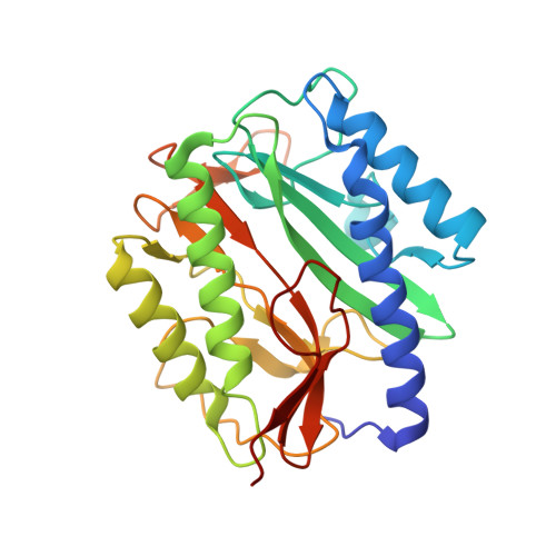
Structural analysis of inhibition of E. coli methionine aminopeptidase: implication of loop flexibility in selective inhibition of bacterial enzymes.
Ma, Z.Q., Xie, S.X., Huang, Q.Q., Nan, F.J., Hurley, T.D., Ye, Q.Z.(2007) BMC Struct Biol 7: 84-84
- PubMed: 18093325
- DOI: https://doi.org/10.1186/1472-6807-7-84
- Primary Citation of Related Structures:
2Q92, 2Q93, 2Q94, 2Q95, 2Q96 - PubMed Abstract:
Methionine aminopeptidase is a potential target of future antibacterial and anticancer drugs. Structural analysis of complexes of the enzyme with its inhibitors provides valuable information for structure-based drug design efforts. Five new X-ray structures of such enzyme-inhibitor complexes were obtained. Analysis of these and other three similar structures reveals the adaptability of a surface-exposed loop bearing Y62, H63, G64 and Y65 (the YHGY loop) that is an integral part of the substrate and inhibitor binding pocket. This adaptability is important for accommodating inhibitors with variations in size. When compared with the human isozymes, this loop either becomes buried in the human type I enzyme due to an N-terminal extension that covers its position or is replaced by a unique insert in the human type II enzyme. The adaptability of the YHGY loop in E. coli methionine aminopeptidase, and likely in other bacterial methionine aminopeptidases, enables the enzyme active pocket to accommodate inhibitors of differing size. The differences in this adaptable loop between the bacterial and human methionine aminopeptidases is a structural feature that can be exploited to design inhibitors of bacterial methionine aminopeptidases as therapeutic agents with minimal inhibition of the corresponding human enzymes.
Organizational Affiliation:
High Throughput Screening Laboratory, University of Kansas, Lawrence, Kansas 66047, USA. zeqiang.ma@vanderbilt.edu


