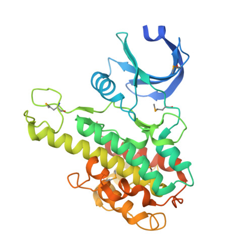
2QR7

2.0A X-ray structure of C-terminal kinase domain of p90 ribosomal S6 kinase 2: Se-Met derivative


Experimental Data Snapshot

  • Method: X-RAY DIFFRACTION
  • Resolution: 2.00 Å
  • R-Value Free: 0.238 
  • R-Value Work: 0.203 
  • R-Value Observed: 0.204 

wwPDB Validation   3D Report Full Report


This is version 1.3 of the entry. See complete history


Literature

Structural basis for activation of the autoinhibitory C-terminal kinase domain of p90 RSK2.

Malakhova, M.Tereshko, V.Lee, S.Y.Yao, K.Cho, Y.-Y.Bode, A.Dong, Z.

(2008) Nat Struct Mol Biol 15: 112-113

  • DOI: https://doi.org/10.1038/nsmb1347
  • Primary Citation of Related Structures:  
    2QR7, 2QR8

  • PubMed Abstract: 

    The X-ray structure at 2.0-A resolution of the p90 ribosomal S6 kinase 2 C-terminal kinase domain revealed a C-terminal autoinhibitory alphaL-helix that was embedded in the kinase scaffold and determines the inactive kinase conformation. We suggest a mechanism of activation through displacement of the alphaL-helix and rearrangement of the conserved residue Glu500, as well as the reorganization of the T-loop into the active conformation.


  • Organizational Affiliation

    The Hormel Institute, University of Minnesota, 801 16th Avenue NE, Austin, Minnesota 55912, USA.


Macromolecules
Find similar proteins by:  (by identity cutoff)  |  3D Structure
Entity ID: 1
MoleculeChains Sequence LengthOrganismDetailsImage
Ribosomal protein S6 kinase alpha-3342Mus musculusMutation(s): 1 
Gene Names: Rps6ka3Rps6ka-rs1Rsk2
EC: 2.7.11.1
UniProt
Find proteins for P18654 (Mus musculus)
Explore P18654 
Go to UniProtKB:  P18654
Entity Groups  
Sequence Clusters30% Identity50% Identity70% Identity90% Identity95% Identity100% Identity
UniProt GroupP18654
Sequence Annotations
Expand
  • Reference Sequence
Small Molecules
Ligands 1 Unique
IDChains Name / Formula / InChI Key2D Diagram3D Interactions
NA
Query on NA

Download Ideal Coordinates CCD File 
B [auth A]SODIUM ION
Na
FKNQFGJONOIPTF-UHFFFAOYSA-N
Modified Residues  1 Unique
IDChains TypeFormula2D DiagramParent
MSE
Query on MSE
A
L-PEPTIDE LINKINGC5 H11 N O2 SeMET
Experimental Data & Validation

Experimental Data

  • Method: X-RAY DIFFRACTION
  • Resolution: 2.00 Å
  • R-Value Free: 0.238 
  • R-Value Work: 0.203 
  • R-Value Observed: 0.204 
  • Space Group: P 41 21 2
Unit Cell:
Length ( Å )Angle ( ˚ )
a = 46.585α = 90
b = 46.585β = 90
c = 293.99γ = 90
Software Package:
Software NamePurpose
REFMACrefinement
HKL-2000data collection
HKL-2000data reduction
HKL-2000data scaling
SOLVEphasing

Structure Validation

View Full Validation Report


Entry History 

Deposition Data

Revision History  (Full details and data files)

  • Version 1.0: 2007-12-11
    Type: Initial release
  • Version 1.1: 2011-07-13
    Changes: Advisory, Version format compliance
  • Version 1.2: 2021-10-20
    Changes: Database references, Derived calculations
  • Version 1.3: 2024-10-30
    Changes: Data collection, Structure summary