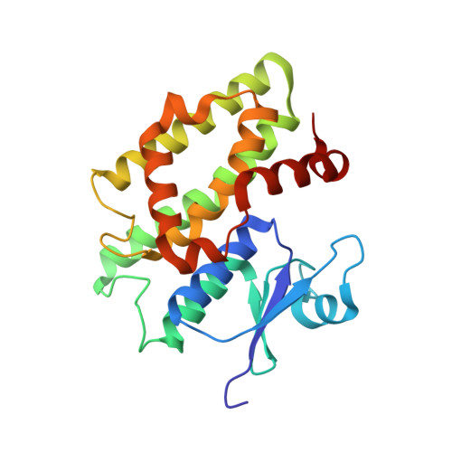
Crystallographic and Functional Characterization of the Fluorodifen-Inducible Glutathione Transferase from Glycine Max Reveals an Active Site Topography Suited for Diphenylether Herbicides and a Novel L-Site.
Axarli, I., Dhavala, P., Papageorgiou, A.C., Labrou, N.E.(2009) J Mol Biol 385: 984
- PubMed: 19014949
- DOI: https://doi.org/10.1016/j.jmb.2008.10.084
- Primary Citation of Related Structures:
2VO4 - PubMed Abstract:
Glutathione transferases (GSTs) from the tau class (GSTU) are unique to plants and have important roles in stress tolerance and the detoxification of herbicides in crops and weeds. A fluorodifen-induced GST isoezyme (GmGSTU4-4) belonging to the tau class was purified from Glycine max by affinity chromatography. This isoenzyme was cloned and expressed in Escherichia coli, and its structural and catalytic properties were investigated. The structure of GmGSTU4-4 was determined at 1.75 A resolution in complex with S-(p-nitrobenzyl)-glutathione (Nb-GSH). The enzyme adopts the canonical GST fold but with a number of functionally important differences. Compared with other plant GSTs, the three-dimensional structure of GmGSTU4-4 primarily shows structural differences in the hydrophobic substrate binding site, the linker segment and the C-terminal region. The X-ray structure identifies key amino acid residues in the hydrophobic binding site (H-site) and provides insights into the substrate specificity and catalytic mechanism of the enzyme. The isoenzyme was highly active in conjugating the diphenylether herbicide fluorodifen. A possible reaction pathway involving the conjugation of glutathione with fluorodifen is described based on site-directed mutagenesis and molecular modeling studies. A serine residue (Ser13) is present in the active site, at a position that would allow it to stabilise the thiolate anion of glutathione and enhance its nucleophilicity. Tyr107 and Arg111 present in the active site are important structural moieties that modulate the catalytic efficiency and specificity of the enzyme, and participate in k(cat) regulation by affecting the rate-limiting step of the catalytic reaction. A hitherto undescribed ligand-binding site (L-site) located in a surface pocket of the enzyme was also found. This site is formed by conserved residues, suggesting it may have an important functional role in the transfer and delivery of bound ligands, presumably to specific protein receptors.
Organizational Affiliation:
Laboratory of Enzyme Technology, Department of Agricultural Biotechnology, Agricultural University of Athens, 75 Iera Odos Street, GR-11855-Athens, Greece.


