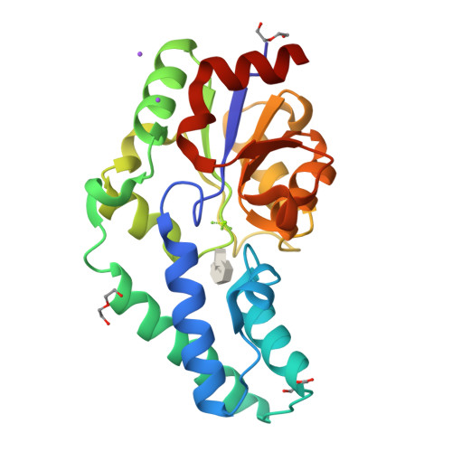
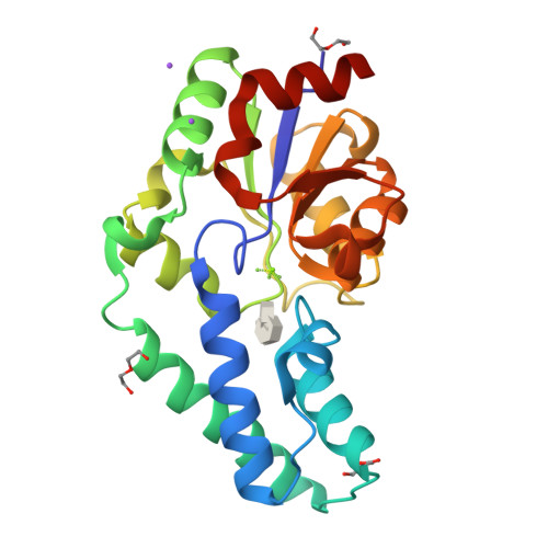
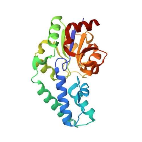
2WF8

Structure of Beta-Phosphoglucomutase inhibited with Glucose-6- phosphate, Glucose-1-phosphate and Beryllium trifluoride


Experimental Data Snapshot

  • Method: X-RAY DIFFRACTION
  • Resolution: 1.20 Å
  • R-Value Free: 0.191 
  • R-Value Work: 0.162 
  • R-Value Observed: 0.163 

Starting Model: experimental
View more details

wwPDB Validation   3D Report Full Report


Ligand Structure Quality Assessment 


This is version 4.2 of the entry. See complete history


Literature

Near attack conformers dominate beta-phosphoglucomutase complexes where geometry and charge distribution reflect those of substrate.

Griffin, J.L.Bowler, M.W.Baxter, N.J.Leigh, K.N.Dannatt, H.R.Hounslow, A.M.Blackburn, G.M.Webster, C.E.Cliff, M.J.Waltho, J.P.

(2012) Proc Natl Acad Sci U S A 109: 6910-6915

  • DOI: https://doi.org/10.1073/pnas.1116855109
  • Primary Citation of Related Structures:  
    2WF8, 2WF9, 2WFA

  • PubMed Abstract: 

    Experimental observations of fluoromagnesate and fluoroaluminate complexes of β-phosphoglucomutase (β-PGM) have demonstrated the importance of charge balance in transition-state stabilization for phosphoryl transfer enzymes. Here, direct observations of ground-state analog complexes of β-PGM involving trifluoroberyllate establish that when the geometry and charge distribution closely match those of the substrate, the distribution of conformers in solution and in the crystal predominantly places the reacting centers in van der Waals proximity. Importantly, two variants are found, both of which satisfy the criteria for near attack conformers. In one variant, the aspartate general base for the reaction is remote from the nucleophile. The nucleophile remains protonated and forms a nonproductive hydrogen bond to the phosphate surrogate. In the other variant, the general base forms a hydrogen bond to the nucleophile that is now correctly orientated for the chemical transfer step. By contrast, in the absence of substrate, the solvent surrounding the phosphate surrogate is arranged to disfavor nucleophilic attack by water. Taken together, the trifluoroberyllate complexes of β-PGM provide a picture of how the enzyme is able to organize itself for the chemical step in catalysis through the population of intermediates that respond to increasing proximity of the nucleophile. These experimental observations show how the enzyme is capable of stabilizing the reaction pathway toward the transition state and also of minimizing unproductive catalysis of aspartyl phosphate hydrolysis.


  • Organizational Affiliation

    Department of Molecular Biology and Biotechnology, Krebs Institute, University of Sheffield, Sheffield S10 2TN, United Kingdom.


Macromolecules
Find similar proteins by:  (by identity cutoff)  |  3D Structure
Entity ID: 1
MoleculeChains Sequence LengthOrganismDetailsImage
BETA-PHOSPHOGLUCOMUTASE221Lactococcus lactisMutation(s): 0 
EC: 5.4.2.6
UniProt
Find proteins for P71447 (Lactococcus lactis subsp. lactis (strain IL1403))
Explore P71447 
Go to UniProtKB:  P71447
Entity Groups  
Sequence Clusters30% Identity50% Identity70% Identity90% Identity95% Identity100% Identity
UniProt GroupP71447
Sequence Annotations
Expand
  • Reference Sequence
Small Molecules
Ligands 6 Unique
IDChains Name / Formula / InChI Key2D Diagram3D Interactions
BG6
Query on BG6

Download Ideal Coordinates CCD File 
E [auth A]6-O-phosphono-beta-D-glucopyranose
C6 H13 O9 P
NBSCHQHZLSJFNQ-VFUOTHLCSA-N
G1P
Query on G1P

Download Ideal Coordinates CCD File 
D [auth A]1-O-phosphono-alpha-D-glucopyranose
C6 H13 O9 P
HXXFSFRBOHSIMQ-VFUOTHLCSA-N
PEG
Query on PEG

Download Ideal Coordinates CCD File 
H [auth A],
I [auth A],
J [auth A]
DI(HYDROXYETHYL)ETHER
C4 H10 O3
MTHSVFCYNBDYFN-UHFFFAOYSA-N
BEF
Query on BEF

Download Ideal Coordinates CCD File 
B [auth A]BERYLLIUM TRIFLUORIDE ION
Be F3
OGIAHMCCNXDTIE-UHFFFAOYSA-K
MG
Query on MG

Download Ideal Coordinates CCD File 
C [auth A]MAGNESIUM ION
Mg
JLVVSXFLKOJNIY-UHFFFAOYSA-N
NA
Query on NA

Download Ideal Coordinates CCD File 
F [auth A],
G [auth A]
SODIUM ION
Na
FKNQFGJONOIPTF-UHFFFAOYSA-N
Experimental Data & Validation

Experimental Data

  • Method: X-RAY DIFFRACTION
  • Resolution: 1.20 Å
  • R-Value Free: 0.191 
  • R-Value Work: 0.162 
  • R-Value Observed: 0.163 
  • Space Group: P 21 21 21
Unit Cell:
Length ( Å )Angle ( ˚ )
a = 37.3α = 90
b = 54.3β = 90
c = 104.2γ = 90
Software Package:
Software NamePurpose
REFMACrefinement
MOSFLMdata reduction
SCALAdata scaling
MOLREPphasing

Structure Validation

View Full Validation Report


Ligand Structure Quality Assessment 


Entry History 

Revision History  (Full details and data files)

  • Version 1.0: 2010-05-26
    Type: Initial release
  • Version 1.1: 2011-09-21
    Changes: Atomic model, Derived calculations, Non-polymer description, Other, Version format compliance
  • Version 1.2: 2012-05-16
    Changes: Other
  • Version 2.0: 2019-01-30
    Changes: Advisory, Atomic model, Data collection, Database references, Experimental preparation
  • Version 3.0: 2019-05-22
    Changes: Advisory, Atomic model, Data collection
  • Version 4.0: 2019-10-02
    Changes: Atomic model, Data collection, Other
  • Version 4.1: 2020-07-29
    Type: Remediation
    Reason: Carbohydrate remediation
    Changes: Data collection, Derived calculations, Structure summary
  • Version 4.2: 2023-12-13
    Changes: Data collection, Database references, Refinement description, Structure summary