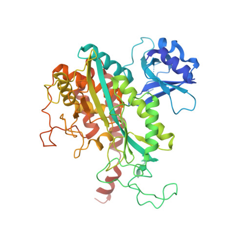
2WGS

Crystal structure of Mycobacterium Tuberculosis Glutamine Synthetase in complex with a purine analogue inhibitor.


Experimental Data Snapshot

  • Method: X-RAY DIFFRACTION
  • Resolution: 2.55 Å
  • R-Value Free: 0.246 
  • R-Value Work: 0.230 
  • R-Value Observed: 0.231 

Starting Model: experimental
View more details

wwPDB Validation   3D Report Full Report


Ligand Structure Quality Assessment 


This is version 1.5 of the entry. See complete history


Literature

Structural Basis for the Inhibition of Mycobacterium Tuberculosis Glutamine Synthetase by Novel ATP-Competitive Inhibitors.

Nilsson, M.T.Krajewski, W.W.Yellagunda, S.Prabhumurthy, S.Chamarahally, G.N.Siddamadappa, C.Srinivasa, B.R.Yahiaoui, S.Larhed, M.Karlen, A.Jones, T.A.Mowbray, S.L.

(2009) J Mol Biol 393: 504

  • DOI: https://doi.org/10.1016/j.jmb.2009.08.028
  • Primary Citation of Related Structures:  
    2WGS, 2WHI

  • PubMed Abstract: 

    Glutamine synthetase (GS, EC 6.3.1.2; also known as gamma-glutamyl:ammonia ligase) catalyzes the ATP-dependent condensation of glutamate and ammonia to form glutamine. The enzyme has essential roles in different tissues and species, which have led to its consideration as a drug or an herbicide target. In this article, we describe studies aimed at the discovery of new antimicrobial agents targeting Mycobacterium tuberculosis, the causative pathogen of tuberculosis. A number of distinct classes of GS inhibitors with an IC(50) of micromolar value or better were identified via high-throughput screening. A commercially available purine analogue similar to one of the clusters identified (the diketopurines), 1-[(3,4-dichlorophenyl)methyl]-3,7-dimethyl-8-morpholin-4-yl-purine-2,6-dione, was also shown to inhibit the enzyme, with a measured IC(50) of 2.5+/-0.4 microM. Two X-ray structures are presented: one is a complex of the enzyme with the purine analogue alone (2.55-A resolution), and the other includes the compound together with methionine sulfoximine phosphate, magnesium and phosphate (2.2-A resolution). The former represents a relaxed, inactive conformation of the enzyme, while the latter is a taut, active one. These structures show that the compound binds at the same position in the nucleotide site, regardless of the conformational state. The ATP-binding site of the human enzyme differs substantially, explaining why it has an approximately 60-fold lower affinity for this compound than the bacterial GS. As part of this work, we devised a new synthetic procedure for generating l-(SR)-methionine sulfoximine phosphate from l-(SR)-methionine sulfoximine, which will facilitate future investigations of novel GS inhibitors.


  • Organizational Affiliation

    Department of Cell and Molecular Biology, Uppsala University, Biomedical Center, Sweden.


Macromolecules
Find similar proteins by:  (by identity cutoff)  |  3D Structure
Entity ID: 1
MoleculeChains Sequence LengthOrganismDetailsImage
GLUTAMINE SYNTHETASE 1
A, B, C, D, E
A, B, C, D, E, F, G, H, I, J, K, L
486Mycobacterium tuberculosis H37RvMutation(s): 0 
EC: 6.3.1.2
UniProt
Find proteins for P9WN39 (Mycobacterium tuberculosis (strain ATCC 25618 / H37Rv))
Explore P9WN39 
Go to UniProtKB:  P9WN39
Entity Groups  
Sequence Clusters30% Identity50% Identity70% Identity90% Identity95% Identity100% Identity
UniProt GroupP9WN39
Sequence Annotations
Expand
  • Reference Sequence
Small Molecules
Ligands 2 Unique
IDChains Name / Formula / InChI Key2D Diagram3D Interactions
1AZ
Query on 1AZ

Download Ideal Coordinates CCD File 
AA [auth H]
CA [auth I]
EA [auth J]
GA [auth K]
IA [auth L]
AA [auth H],
CA [auth I],
EA [auth J],
GA [auth K],
IA [auth L],
M [auth A],
O [auth B],
Q [auth C],
S [auth D],
U [auth E],
W [auth F],
Y [auth G]
1-(3,4-dichlorobenzyl)-3,7-dimethyl-8-morpholin-4-yl-3,7-dihydro-1H-purine-2,6-dione
C18 H19 Cl2 N5 O3
BFRVAZYYGGLHQS-UHFFFAOYSA-N
CL
Query on CL

Download Ideal Coordinates CCD File 
BA [auth H]
DA [auth I]
FA [auth J]
HA [auth K]
JA [auth L]
BA [auth H],
DA [auth I],
FA [auth J],
HA [auth K],
JA [auth L],
N [auth A],
P [auth B],
R [auth C],
T [auth D],
V [auth E],
X [auth F],
Z [auth G]
CHLORIDE ION
Cl
VEXZGXHMUGYJMC-UHFFFAOYSA-M
Binding Affinity Annotations 
IDSourceBinding Affinity
1AZ PDBBind:  2WGS IC50: 2500 (nM) from 1 assay(s)
Experimental Data & Validation

Experimental Data

  • Method: X-RAY DIFFRACTION
  • Resolution: 2.55 Å
  • R-Value Free: 0.246 
  • R-Value Work: 0.230 
  • R-Value Observed: 0.231 
  • Space Group: P 21 21 21
Unit Cell:
Length ( Å )Angle ( ˚ )
a = 134.95α = 90
b = 203.18β = 90
c = 230.2γ = 90
Software Package:
Software NamePurpose
REFMACrefinement
XDSdata reduction
XSCALEdata scaling
MOLREPphasing

Structure Validation

View Full Validation Report


Ligand Structure Quality Assessment 


Entry History 

Deposition Data

Revision History  (Full details and data files)

  • Version 1.0: 2009-09-01
    Type: Initial release
  • Version 1.1: 2011-05-08
    Changes: Version format compliance
  • Version 1.2: 2011-07-13
    Changes: Version format compliance
  • Version 1.3: 2019-05-08
    Changes: Data collection, Experimental preparation, Other
  • Version 1.4: 2019-05-15
    Changes: Data collection, Experimental preparation
  • Version 1.5: 2023-12-13
    Changes: Data collection, Database references, Derived calculations, Other, Refinement description