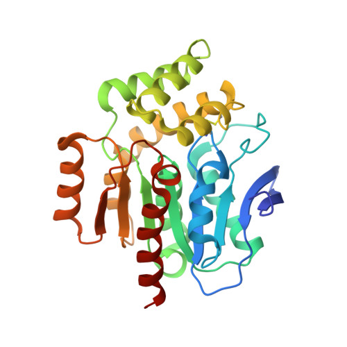
2WM2

CRYSTAL STRUCTURE OF THE COFACTOR-DEVOID 1-H-3-HYDROXY-4- OXOQUINALDINE 2,4-DIOXYGENASE (HOD) FROM ARTHROBACTER NITROGUAJACOLICUS RU61A IN COMPLEX WITH CHLORIDE


Experimental Data Snapshot

  • Method: X-RAY DIFFRACTION
  • Resolution: 2.70 Å
  • R-Value Free: 0.246 
  • R-Value Work: 0.211 
  • R-Value Observed: 0.213 

wwPDB Validation   3D Report Full Report


Ligand Structure Quality Assessment 


This is version 1.3 of the entry. See complete history


Literature

Structural Basis for Cofactor-Independent Dioxygenation of N-Heteroaromatic Compounds at the {Alpha}/{Beta}-Hydrolase Fold.

Steiner, R.A.Janssen, H.J.Roversi, P.Oakley, A.J.Fetzner, S.

(2010) Proc Natl Acad Sci U S A 107: 657

  • DOI: https://doi.org/10.1073/pnas.0909033107
  • Primary Citation of Related Structures:  
    2WJ3, 2WJ4, 2WJ6, 2WM2, 3IBT

  • PubMed Abstract: 

    Enzymatic catalysis of oxygenation reactions in the absence of metal or organic cofactors is a considerable biochemical challenge. The CO-forming 1-H-3-hydroxy-4-oxoquinaldine 2,4-dioxygenase (HOD) from Arthrobacter nitroguajacolicus Rü61a and 1-H-3-hydroxy-4-oxoquinoline 2,4-dioxygenase (QDO) from Pseudomonas putida 33/1 are homologous cofactor-independent dioxygenases involved in the breakdown of N-heteroaromatic compounds. To date, they are the only dioxygenases suggested to belong to the alpha/beta-hydrolase fold superfamily. Members of this family typically catalyze hydrolytic processes rather than oxygenation reactions. We present here the crystal structures of both HOD and QDO in their native state as well as the structure of HOD in complex with its natural 1-H-3-hydroxy-4-oxoquinaldine substrate, its N-acetylanthranilate reaction product, and chloride as dioxygen mimic. HOD and QDO are structurally very similar. They possess a classical alpha/beta-hydrolase fold core domain additionally equipped with a cap domain. Organic substrates bind in a preorganized active site with an orientation ideally suited for selective deprotonation of their hydroxyl group by a His/Asp charge-relay system affording the generation of electron-donating species. The "oxyanion hole" of the alpha/beta-hydrolase fold, typically employed to stabilize the tetrahedral intermediate in ester hydrolysis reactions, is utilized here to host and control oxygen chemistry, which is proposed to involve a peroxide anion intermediate. Product release by proton back transfer from the catalytic histidine is driven by minimization of intramolecular charge repulsion. Structural and kinetic data suggest a nonnucleophilic general-base mechanism. Our analysis provides a framework to explain cofactor-independent dioxygenation within a protein architecture generally employed to catalyze hydrolytic reactions.


  • Organizational Affiliation

    Randall Division of Cell and Molecular Biophysics, King's College London, New Hunt's House, Guy's Campus, London SE1 1UL, England. roberto.steiner@kcl.ac.uk


Macromolecules
Find similar proteins by:  (by identity cutoff)  |  3D Structure
Entity ID: 1
MoleculeChains Sequence LengthOrganismDetailsImage
1-H-3-HYDROXY-4-OXOQUINALDINE 2,4-DIOXYGENASE
A, B, C, D
279Paenarthrobacter nitroguajacolicusMutation(s): 1 
EC: 1.13.11.48
UniProt
Find proteins for O31266 (Paenarthrobacter nitroguajacolicus)
Explore O31266 
Go to UniProtKB:  O31266
Entity Groups  
Sequence Clusters30% Identity50% Identity70% Identity90% Identity95% Identity100% Identity
UniProt GroupO31266
Sequence Annotations
Expand
  • Reference Sequence
Small Molecules
Ligands 5 Unique
IDChains Name / Formula / InChI Key2D Diagram3D Interactions
SRT
Query on SRT

Download Ideal Coordinates CCD File 
F [auth A]
HA [auth C]
N [auth A]
O [auth A]
P [auth A]
F [auth A],
HA [auth C],
N [auth A],
O [auth A],
P [auth A],
X [auth B],
Y [auth B]
S,R MESO-TARTARIC ACID
C4 H6 O6
FEWJPZIEWOKRBE-XIXRPRMCSA-N
GOL
Query on GOL

Download Ideal Coordinates CCD File 
AA [auth C]
E [auth A]
G [auth A]
IA [auth D]
JA [auth D]
AA [auth C],
E [auth A],
G [auth A],
IA [auth D],
JA [auth D],
Q [auth B],
R [auth B],
Z [auth C]
GLYCEROL
C3 H8 O3
PEDCQBHIVMGVHV-UHFFFAOYSA-N
K
Query on K

Download Ideal Coordinates CCD File 
BA [auth C],
H [auth A],
KA [auth D],
S [auth B]
POTASSIUM ION
K
NPYPAHLBTDXSSS-UHFFFAOYSA-N
CL
Query on CL

Download Ideal Coordinates CCD File 
GA [auth C]
J [auth A]
K [auth A]
L [auth A]
M [auth A]
GA [auth C],
J [auth A],
K [auth A],
L [auth A],
M [auth A],
NA [auth D],
QA [auth D],
W [auth B]
CHLORIDE ION
Cl
VEXZGXHMUGYJMC-UHFFFAOYSA-M
NA
Query on NA

Download Ideal Coordinates CCD File 
CA [auth C]
DA [auth C]
EA [auth C]
FA [auth C]
I [auth A]
CA [auth C],
DA [auth C],
EA [auth C],
FA [auth C],
I [auth A],
LA [auth D],
MA [auth D],
OA [auth D],
PA [auth D],
T [auth B],
U [auth B],
V [auth B]
SODIUM ION
Na
FKNQFGJONOIPTF-UHFFFAOYSA-N
Experimental Data & Validation

Experimental Data

  • Method: X-RAY DIFFRACTION
  • Resolution: 2.70 Å
  • R-Value Free: 0.246 
  • R-Value Work: 0.211 
  • R-Value Observed: 0.213 
  • Space Group: P 21 21 21
Unit Cell:
Length ( Å )Angle ( ˚ )
a = 45.77α = 90
b = 167β = 90
c = 166.94γ = 90
Software Package:
Software NamePurpose
REFMACrefinement
MOSFLMdata reduction
SCALAdata scaling

Structure Validation

View Full Validation Report


Ligand Structure Quality Assessment 


Entry History 

Deposition Data

Revision History  (Full details and data files)

  • Version 1.0: 2010-01-26
    Type: Initial release
  • Version 1.1: 2011-05-08
    Changes: Version format compliance
  • Version 1.2: 2011-07-13
    Changes: Version format compliance
  • Version 1.3: 2024-11-20
    Changes: Data collection, Database references, Derived calculations, Other, Refinement description, Structure summary