2XOW

Structure of GlpG in complex with a mechanism-based isocoumarin inhibitor


Experimental Data Snapshot

  • Method: X-RAY DIFFRACTION
  • Resolution: 2.09 Å
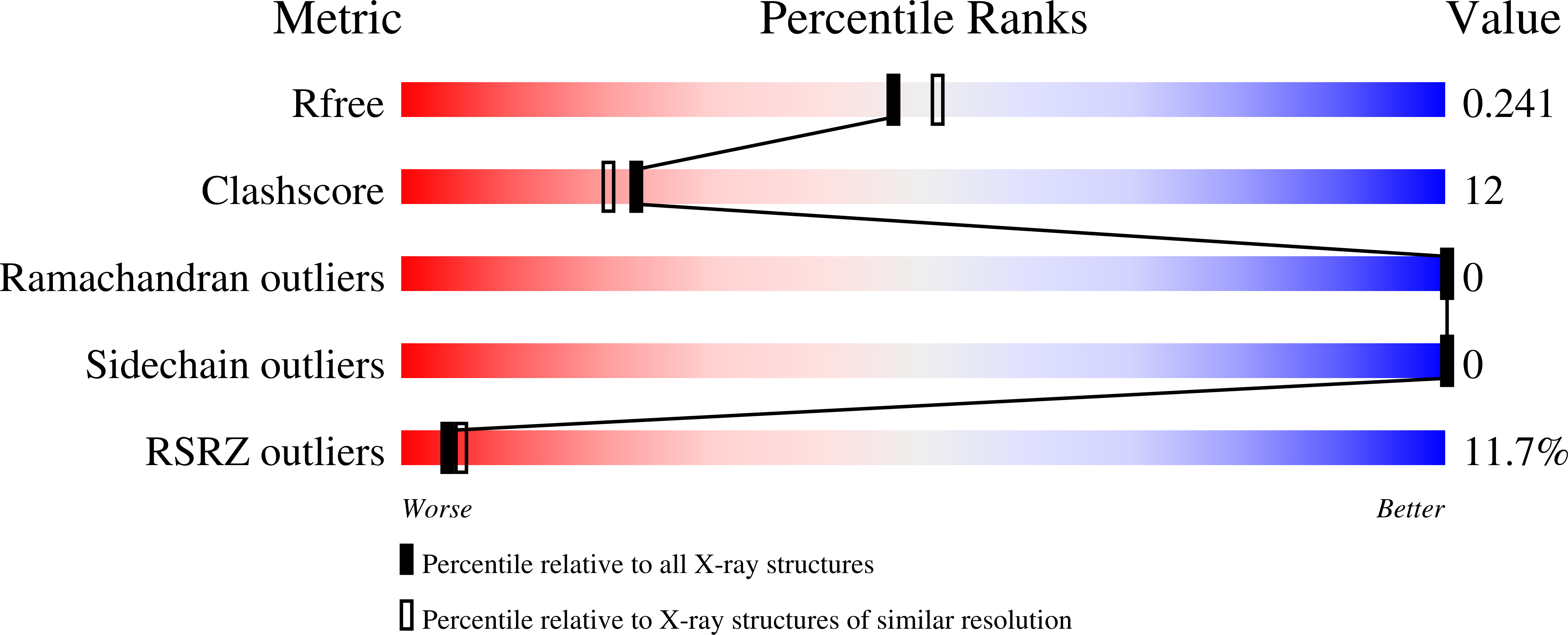
  • R-Value Free: 0.243 
  • R-Value Work: 0.198 
  • R-Value Observed: 0.200 

Starting Model: experimental
View more details

wwPDB Validation   3D Report Full Report


Ligand Structure Quality Assessment 


This is version 1.5 of the entry. See complete history


Literature

The Structural Basis for Catalysis and Substrate Specificity of a Rhomboid Protease

Vinothkumar, K.R.Strisovsky, K.Andreeva, A.Christova, Y.Verhelst, S.Freeman, M.

(2010) EMBO J 29: 3797

  • DOI: https://doi.org/10.1038/emboj.2010.243
  • Primary Citation of Related Structures:  
    2XOV, 2XOW

  • PubMed Abstract: 

    Rhomboids are intramembrane proteases that use a catalytic dyad of serine and histidine for proteolysis. They are conserved in both prokaryotes and eukaryotes and regulate cellular processes as diverse as intercellular signalling, parasitic invasion of host cells, and mitochondrial morphology. Their widespread biological significance and consequent medical potential provides a strong incentive to understand the mechanism of these unusual enzymes for identification of specific inhibitors. In this study, we describe the structure of Escherichia coli rhomboid GlpG covalently bound to a mechanism-based isocoumarin inhibitor. We identify the position of the oxyanion hole, and the S₁- and S₂'-binding subsites of GlpG, which are the key determinants of substrate specificity. The inhibitor-bound structure suggests that subtle structural change is sufficient for catalysis, as opposed to large changes proposed from previous structures of unliganded GlpG. Using bound inhibitor as a template, we present a model for substrate binding at the active site and biochemically test its validity. This study provides a foundation for a structural explanation of rhomboid specificity and mechanism, and for inhibitor design.


  • Organizational Affiliation

    MRC Laboratory of Molecular Biology, Cambridge, UK. vkumar@mrc-lmb.cam.ac.uk


Macromolecules
Find similar proteins by:  (by identity cutoff)  |  3D Structure
Entity ID: 1
MoleculeChains Sequence LengthOrganismDetailsImage
RHOMBOID PROTEASE GLPG179Escherichia coliMutation(s): 0 
EC: 3.4.21.105
Membrane Entity: Yes 
UniProt
Find proteins for P09391 (Escherichia coli (strain K12))
Explore P09391 
Go to UniProtKB:  P09391
Entity Groups  
Sequence Clusters30% Identity50% Identity70% Identity90% Identity95% Identity100% Identity
UniProt GroupP09391
Sequence Annotations
Expand
  • Reference Sequence
Small Molecules
Ligands 2 Unique
IDChains Name / Formula / InChI Key2D Diagram3D Interactions
BNG
Query on BNG

Download Ideal Coordinates CCD File 
C [auth A]
D [auth A]
E [auth A]
F [auth A]
G [auth A]
C [auth A],
D [auth A],
E [auth A],
F [auth A],
G [auth A],
H [auth A],
I [auth A],
J [auth A],
K [auth A],
L [auth A],
M [auth A],
N [auth A],
O [auth A],
P [auth A],
Q [auth A],
R [auth A]
nonyl beta-D-glucopyranoside
C15 H30 O6
QFAPUKLCALRPLH-UXXRCYHCSA-N
ISM
Query on ISM

Download Ideal Coordinates CCD File 
B [auth A]5-AMINO-2-(2-METHOXY-2-OXOETHYL)BENZOIC ACID
C10 H11 N O4
NRWKEWXVSUGTJF-UHFFFAOYSA-N
Binding Affinity Annotations 
IDSourceBinding Affinity
ISM PDBBind:  2XOW IC50: 6000 (nM) from 1 assay(s)
Experimental Data & Validation

Experimental Data

  • Method: X-RAY DIFFRACTION
  • Resolution: 2.09 Å
  • R-Value Free: 0.243 
  • R-Value Work: 0.198 
  • R-Value Observed: 0.200 
  • Space Group: H 3 2
Unit Cell:
Length ( Å )Angle ( ˚ )
a = 110.69α = 90
b = 110.69β = 90
c = 122.151γ = 120
Software Package:
Software NamePurpose
PHENIXrefinement
MOSFLMdata reduction
SCALAdata scaling
PHASERphasing

Structure Validation

View Full Validation Report


Ligand Structure Quality Assessment 


Entry History 

Deposition Data

Revision History  (Full details and data files)

  • Version 1.0: 2010-10-13
    Type: Initial release
  • Version 1.1: 2011-05-26
    Changes: Version format compliance
  • Version 1.2: 2011-07-13
    Changes: Version format compliance
  • Version 1.3: 2019-02-27
    Changes: Data collection, Derived calculations, Experimental preparation, Other
  • Version 1.4: 2020-07-29
    Type: Remediation
    Reason: Carbohydrate remediation
    Changes: Data collection, Derived calculations, Other, Structure summary
  • Version 1.5: 2023-12-20
    Changes: Data collection, Database references, Refinement description, Structure summary