Substitution of Glu122 by glutamine revealed the function of the second water molecule as a proton donor in the binuclear metal enzyme creatininase
Yamashita, K., Nakajima, Y., Matsushita, H., Nishiya, Y., Yamazawa, R., Wu, Y.F., Matsubara, F., Oyama, H., Ito, K., Yoshimoto, T.(2010) J Mol Biol 396: 1081-1096
- PubMed: 20043918
- DOI: https://doi.org/10.1016/j.jmb.2009.12.045
- Primary Citation of Related Structures:
3A6D, 3A6E, 3A6F, 3A6G, 3A6H, 3A6J, 3A6K, 3A6L - PubMed Abstract:
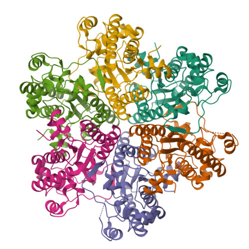
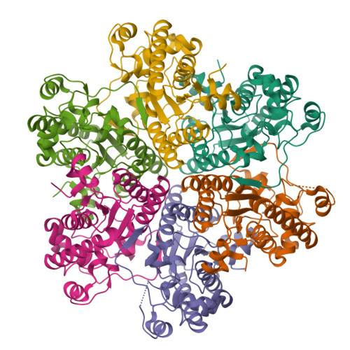
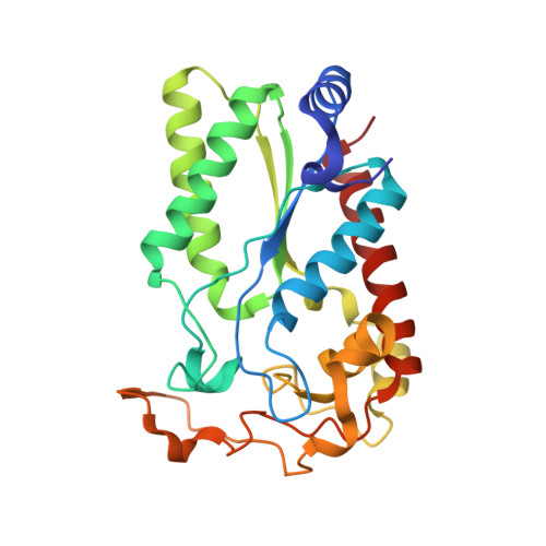
Creatininase is a binuclear zinc enzyme and catalyzes the reversible conversion of creatinine to creatine. It exhibits an open-closed conformational change upon substrate binding, and the differences in the conformations of Tyr121, Trp154, and the loop region containing Trp174 were evident in the enzyme-creatine complex when compared to those in the ligand-free enzyme. We have determined the crystal structure of the enzyme complexed with a 1-methylguanidine. All subunits in the complex existed as the closed form, and the binding mode of creatinine was estimated. Site-directed mutagenesis revealed that the hydrophobic residues that show conformational change upon substrate binding are important for the enzyme activity. We propose a catalytic mechanism of creatininase in which two water molecules have significant roles. The first molecule is a hydroxide ion (Wat1) that is bound as a bridge between the two metal ions and attacks the carbonyl carbon of the substrate. The second molecule is a water molecule (Wat2) that is bound to the carboxyl group of Glu122 and functions as a proton donor in catalysis. The activity of the E122Q mutant was very low and it was only partially restored by the addition of ZnCl(2) or MnCl(2). In the E122Q mutant, k(cat) is drastically decreased, indicating that Glu122 is important for catalysis. X-ray crystallographic study and the atomic absorption spectrometry analysis of the E122Q mutant-substrate complex revealed that the drastic decrease of the activity of the E122Q was caused by not only the loss of one Zn ion at the Metal1 site but also a critical function of Glu122, which most likely exists for a proton transfer step through Wat2.
Organizational Affiliation:
Graduate School of Biomedical Sciences, Nagasaki University, 1-14 Bunkyo-machi, Nagasaki 852-8521, Japan.


