The crystal structure of human Delta4-3-ketosteroid 5beta-reductase defines the functional role of the residues of the catalytic tetrad in the steroid double bond reduction mechanism.
Faucher, F., Cantin, L., Luu-The, V., Labrie, F., Breton, R.(2008) Biochemistry 47: 8261-8270
- PubMed: 18624455
- DOI: https://doi.org/10.1021/bi800572s
- Primary Citation of Related Structures:
3CAV - PubMed Abstract:
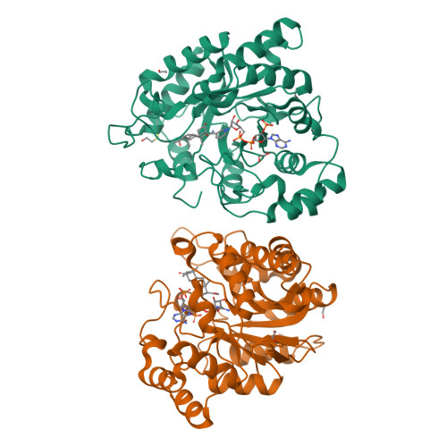
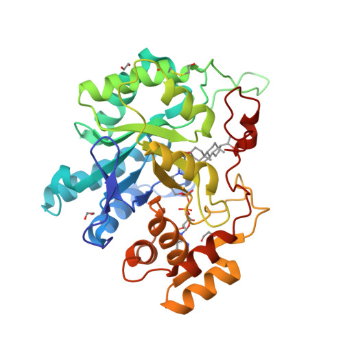
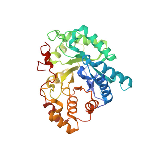
The 5beta-reductases (AKR1D1-3) are unique enzymes able to catalyze efficiently and in a stereospecific manner the 5beta-reduction of the C4-C5 double bond found into Delta4-3-ketosteroids, including steroid hormones and bile acids. Multiple-sequence alignments and mutagenic studies have already identified one of the residues presumably located at their active site, Glu 120, as the major molecular determinant for the unique activity displayed by 5beta-reductases. To define the exact role played by this glutamate in the catalytic activity of these enzymes, biochemical and structural studies on human 5beta-reductase (h5beta-red) have been undertaken. The crystal structure of h5beta-red in a ternary complex with NADP (+) and 5beta-dihydroprogesterone (5beta-DHP), the product of the 5beta-reduction of progesterone (Prog), revealed that Glu 120 does not interact directly with the other catalytic residues, as previously hypothesized, thus suggesting that this residue is not directly involved in catalysis but could instead be important for the proper positioning of the steroid substrate in the catalytic site. On the basis of our structural results, we thus propose a realistic scheme for the catalytic mechanism of the C4-C5 double bond reduction. We also propose that bile acid precursors such as 7alpha-hydroxy-4-cholesten-3-one and 7alpha,12alpha-dihydroxy-4-cholesten-3-one, when bound to the active site of h5beta-red, can establish supplementary contacts with Tyr 26 and Tyr 132, two residues delineating the steroid-binding cavity. These additional contacts very likely account for the higher activity of h5beta-red toward the bile acid intermediates versus steroid hormones. Finally, in light of the structural data now available, we attempt to interpret the likely consequences of mutations already identified in the gene encoding the h5beta-red enzyme which lead to a reduction of its enzymatic activity and which can progress to severe liver function failure.
Organizational Affiliation:
Oncology and Molecular Endocrinology Research Center, Laval University Medical Center, Québec (QC), G1V 4G2 Canada.


