3D4V | pdb_00003d4v

Crystal Structure of an AlkA Host/Guest Complex N7MethylGuanine:Cytosine Base Pair


Experimental Data Snapshot

  • Method: X-RAY DIFFRACTION
  • Resolution: 2.90 Å
  • R-Value Free: 
    0.275 (Depositor) 
  • R-Value Work: 
    0.208 (Depositor), 0.210 (DCC) 

wwPDB Validation   3D Report Full Report


This is version 1.3 of the entry. See complete history


Literature

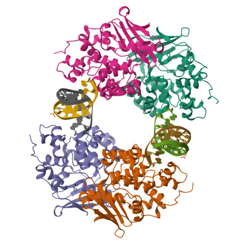
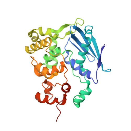
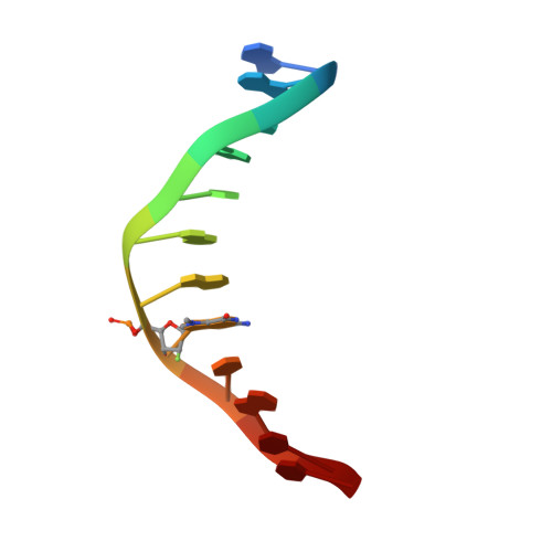
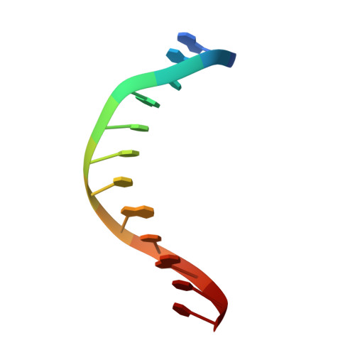
Synthesis and structure of duplex DNA containing the genotoxic nucleobase lesion N7-methylguanine.

Lee, S.Bowman, B.R.Ueno, Y.Wang, S.Verdine, G.L.

(2008) J Am Chem Soc 130: 11570-11571

  • DOI: https://doi.org/10.1021/ja8025328
  • Primary Citation of Related Structures:  
    3D4V

  • PubMed Abstract: 

    The predominant product of aberrant DNA methylation is the genotoxic lesion N7-methyl-2'-deoxyguanosine (m7dG). M7dG is recognized and excised by lesion-specific DNA glycosylases, namely AlkA in E. coli and Aag in humans. Structural studies of m7dG recognition and catalysis by these enzymes have been hampered due to a lack of efficient means by which to incorporate the chemically labile m7dG moiety site-specifically into DNA on a preparative scale. Here we report a solution to this problem. We stabilized the lesion toward acid-catalyzed and glycosylase-catalyzed depurination by 2'-fluorination and toward base-catalyzed degradation using mild, nonaqueous conditions in the DNA deprotection reaction. Duplex DNA containing 2'-fluoro-m7dG (Fm7dG) cocrystallized with AlkA as a host-guest complex in which the lesion-containing segment of DNA was nearly devoid of protein contacts, thus enabling the first direct visualization of the N7-methylguanine lesion nucleobase in DNA. The structure reveals that the base-pairing mode of Fm7dG:C is nearly identical to that of G:C, and Fm7dG does not induce any apparent structural disturbance of the duplex structure. These observations suggest that AlkA and Aag must perform a structurally invasive interrogation of DNA in order to detect the presence of intrahelical m7dG lesions.


  • Organizational Affiliation

    Department of Chemistry and Chemical Biology, Harvard University, Cambridge, Massachusetts 02138, USA.


Macromolecules

Find similar proteins by:  (by identity cutoff)  |  3D Structure
Entity ID: 1
MoleculeChains Sequence LengthOrganismDetailsImage
DNA-3-methyladenine glycosylase 2
A, B, C, D
282Escherichia coliMutation(s): 0 
Gene Names: alkAaidAb2068JW2053
EC: 3.2.2.21
UniProt
Find proteins for P04395 (Escherichia coli (strain K12))
Explore P04395 
Go to UniProtKB:  P04395
Entity Groups  
Sequence Clusters30% Identity50% Identity70% Identity90% Identity95% Identity100% Identity
UniProt GroupP04395
Sequence Annotations
Expand
  • Reference Sequence
Experimental Data & Validation

Experimental Data

  • Method: X-RAY DIFFRACTION
  • Resolution: 2.90 Å
  • R-Value Free:  0.275 (Depositor) 
  • R-Value Work:  0.208 (Depositor), 0.210 (DCC) 
Space Group: P 1 21 1
Unit Cell:
Length ( Å )Angle ( ˚ )
a = 74.1α = 90
b = 100.361β = 94.45
c = 102.281γ = 90
Software Package:
Software NamePurpose
DENZOdata reduction
SCALEPACKdata scaling
CNSrefinement
PDB_EXTRACTdata extraction

Structure Validation

View Full Validation Report


Entry History 

Deposition Data

Revision History  (Full details and data files)

  • Version 1.0: 2008-09-09
    Type: Initial release
  • Version 1.1: 2011-07-13
    Changes: Version format compliance
  • Version 1.2: 2017-10-25
    Changes: Refinement description
  • Version 1.3: 2024-02-21
    Changes: Data collection, Database references, Derived calculations