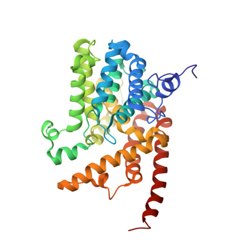
3DYQ

human phosphodiestrase 9 (inhibited by omitting divalent cation) in complex with cGMP


Experimental Data Snapshot

  • Method: X-RAY DIFFRACTION
  • Resolution: 2.50 Å
  • R-Value Free: 0.226 
  • R-Value Work: 0.196 
  • R-Value Observed: 0.199 

Starting Model: experimental
View more details

wwPDB Validation   3D Report Full Report


Ligand Structure Quality Assessment 


This is version 1.2 of the entry. See complete history


Literature

Structural basis for the catalytic mechanism of human phosphodiesterase 9.

Liu, S.Mansour, M.N.Dillman, K.S.Perez, J.R.Danley, D.E.Aeed, P.A.Simons, S.P.Lemotte, P.K.Menniti, F.S.

(2008) Proc Natl Acad Sci U S A 105: 13309-13314

  • DOI: https://doi.org/10.1073/pnas.0708850105
  • Primary Citation of Related Structures:  
    3DY8, 3DYL, 3DYN, 3DYQ, 3DYS

  • PubMed Abstract: 

    The phosphodiesterases (PDEs) are metal ion-dependent enzymes that regulate cellular signaling by metabolic inactivation of the ubiquitous second messengers cAMP and cGMP. In this role, the PDEs are involved in many biological and metabolic processes and are proven targets of successful drugs for the treatments of a wide range of diseases. However, because of the rapidity of the hydrolysis reaction, an experimental knowledge of the enzymatic mechanisms of the PDEs at the atomic level is still lacking. Here, we report the structures of reaction intermediates accumulated at the reaction steady state in PDE9/crystal and preserved by freeze-trapping. These structures reveal the catalytic process of a PDE and explain the substrate specificity of PDE9 in an actual reaction and the cation requirements of PDEs in general.


  • Organizational Affiliation

    Pfizer Global Research and Development, Pfizer Inc., Eastern Point Road, Groton, CT 06340, USA. shenping.liu@pfizer.com


Macromolecules
Find similar proteins by:  (by identity cutoff)  |  3D Structure
Entity ID: 1
MoleculeChains Sequence LengthOrganismDetailsImage
High affinity cGMP-specific 3',5'-cyclic phosphodiesterase 9A
A, B
329Homo sapiensMutation(s): 0 
Gene Names: PDE9A
EC: 3.1.4.35
UniProt & NIH Common Fund Data Resources
Find proteins for O76083 (Homo sapiens)
Explore O76083 
Go to UniProtKB:  O76083
PHAROS:  O76083
GTEx:  ENSG00000160191 
Entity Groups  
Sequence Clusters30% Identity50% Identity70% Identity90% Identity95% Identity100% Identity
UniProt GroupO76083
Sequence Annotations
Expand
  • Reference Sequence
Small Molecules
Binding Affinity Annotations 
IDSourceBinding Affinity
IBM BindingDB:  3DYQ IC50: min: 1.17e+5, max: 2.00e+5 (nM) from 2 assay(s)
Experimental Data & Validation

Experimental Data

  • Method: X-RAY DIFFRACTION
  • Resolution: 2.50 Å
  • R-Value Free: 0.226 
  • R-Value Work: 0.196 
  • R-Value Observed: 0.199 
  • Space Group: P 41 21 2
Unit Cell:
Length ( Å )Angle ( ˚ )
a = 103.65α = 90
b = 103.65β = 90
c = 270.041γ = 90
Software Package:
Software NamePurpose
REFMACrefinement

Structure Validation

View Full Validation Report


Ligand Structure Quality Assessment 


Entry History 

Deposition Data

Revision History  (Full details and data files)

  • Version 1.0: 2008-09-16
    Type: Initial release
  • Version 1.1: 2011-07-13
    Changes: Advisory, Refinement description, Version format compliance
  • Version 1.2: 2023-08-30
    Changes: Data collection, Database references, Derived calculations, Refinement description