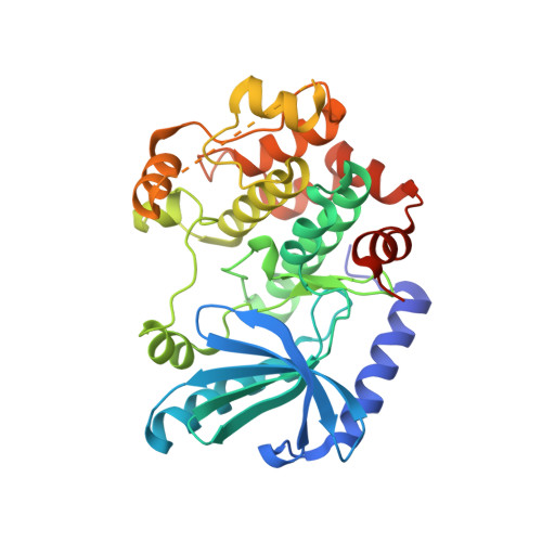
Crystal Structures of MEK1 Binary and Ternary Complexes with Nucleotides and Inhibitors.
Fischmann, T.O., Smith, C.K., Mayhood, T.W., Myers, J.E., Reichert, P., Mannarino, A., Carr, D., Zhu, H., Wong, J., Yang, R.S., Le, H.V., Madison, V.S.(2009) Biochemistry 48: 2661-2674
- PubMed: 19161339
- DOI: https://doi.org/10.1021/bi801898e
- Primary Citation of Related Structures:
3EQC, 3EQD, 3EQF, 3EQG, 3EQH, 3EQI - PubMed Abstract:
MEK1 is a member of the MAPK signal transduction pathway that responds to growth factors and cytokines. We have determined that the kinase domain spans residues 35-382 by proteolytic cleavage. The complete kinase domain has been crystallized and its X-ray crystal structure as a complex with magnesium and ATP-gammaS determined at 2.1 A. Unlike crystals of a truncated kinase domain previously published, the crystals of the intact domain can be grown either as a binary complex with a nucleotide or as a ternary complex with a nucleotide and one of a multitude of allosteric inhibitors. Further, the crystals allow for the determination of costructures with ATP competitive inhibitors. We describe the structures of nonphosphorylated MEK1 (npMEK1) binary complexes with ADP and K252a, an ATP-competitive inhibitor (see Table 1), at 1.9 and 2.7 A resolution, respectively. Ternary complexes have also been solved between npMEK1, a nucleotide, and an allosteric non-ATP competitive inhibitor: ATP-gammaS with compound 1 and ADP with either U0126 or the MEK1 clinical candidate PD325089 at 1.8, 2.0, and 2.5 A, respectively. Compound 1 is structurally similar to PD325901. These structures illustrate fundamental differences among various mechanisms of inhibition at the molecular level. Residues 44-51 have previously been shown to play a negative regulatory role in MEK1 activity. The crystal structure of the integral kinase domain provides a structural rationale for the role of these residues. They form helix A and repress enzymatic activity by stabilizing an inactive conformation in which helix C is displaced from its active state position. Finally, the structure provides for the first time a molecular rationale that explains how mutations in MEK may lead to the cardio-facio-cutaneous syndrome.
Organizational Affiliation:
Schering-Plough Research Institute, Kenilworth, New Jersey 07033, USA. thierry.fischmann@spcorp.com


