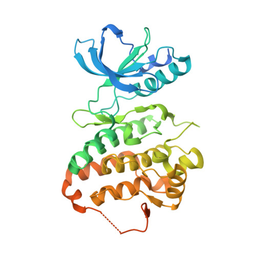
3FY2

Human EphA3 Kinase and Juxtamembrane Region Bound to Substrate KQWDNYEFIW


Experimental Data Snapshot

  • Method: X-RAY DIFFRACTION
  • Resolution: 1.80 Å
  • R-Value Free: 0.212 
  • R-Value Work: 0.179 
  • R-Value Observed: 0.181 

Starting Model: experimental
View more details

wwPDB Validation   3D Report Full Report


This is version 1.5 of the entry. See complete history


Literature

Structural recognition of an optimized substrate for the ephrin family of receptor tyrosine kinases.

Davis, T.L.Walker, J.R.Allali-Hassani, A.Parker, S.A.Turk, B.E.Dhe-Paganon, S.

(2009) FEBS J 276: 4395-4404

  • DOI: https://doi.org/10.1111/j.1742-4658.2009.07147.x
  • Primary Citation of Related Structures:  
    3FXX, 3FY2

  • PubMed Abstract: 

    Ephrin receptor tyrosine kinase A3 (EphA3, EC 2.7.10.1) is a member of a unique branch of the kinome in which downstream signaling occurs in both ligand- and receptor-expressing cells. Consequently, the ephrins and ephrin receptor tyrosine kinases often mediate processes involving cell-cell contact, including cellular adhesion or repulsion, developmental remodeling and neuronal mapping. The receptor is also frequently overexpressed in invasive cancers, including breast, small-cell lung and gastrointestinal cancers. However, little is known about direct substrates of EphA3 kinase and no chemical probes are available. Using a library approach, we found a short peptide sequence that is a good substrate for EphA3 and is suitable for co-crystallization studies. Complex structures show multiple contacts between kinase and substrates; in particular, two residues undergo conformational changes and by mutation are found to be important for substrate binding and turnover. In addition, a difference in catalytic efficiency between EPH kinase family members is observed. These results provide insight into the mechanism of substrate binding to these developmentally integral enzymes.


  • Organizational Affiliation

    Structural Genomics Consortium, University of Toronto, Canada.


Macromolecules
Find similar proteins by:  (by identity cutoff)  |  3D Structure
Entity ID: 1
MoleculeChains Sequence LengthOrganismDetailsImage
Ephrin type-A receptor 3371Homo sapiensMutation(s): 1 
Gene Names: EPHA3ETKETK1HEKTYRO4
EC: 2.7.10.1
UniProt & NIH Common Fund Data Resources
Find proteins for P29320 (Homo sapiens)
Explore P29320 
Go to UniProtKB:  P29320
PHAROS:  P29320
GTEx:  ENSG00000044524 
Entity Groups  
Sequence Clusters30% Identity50% Identity70% Identity90% Identity95% Identity100% Identity
UniProt GroupP29320
Sequence Annotations
Expand
  • Reference Sequence

Find similar proteins by:  Sequence   |   3D Structure  

Entity ID: 2
MoleculeChains Sequence LengthOrganismDetailsImage
peptide substrate10N/AMutation(s): 0 
Entity Groups  
Sequence Clusters30% Identity50% Identity70% Identity90% Identity95% Identity100% Identity
Sequence Annotations
Expand
  • Reference Sequence
Experimental Data & Validation

Experimental Data

  • Method: X-RAY DIFFRACTION
  • Resolution: 1.80 Å
  • R-Value Free: 0.212 
  • R-Value Work: 0.179 
  • R-Value Observed: 0.181 
  • Space Group: P 1 21 1
Unit Cell:
Length ( Å )Angle ( ˚ )
a = 53.824α = 90
b = 38.261β = 102.05
c = 76.365γ = 90
Software Package:
Software NamePurpose
MOSFLMdata reduction
SCALAdata scaling
PHASERphasing
REFMACrefinement
PDB_EXTRACTdata extraction
SBC-Collectdata collection

Structure Validation

View Full Validation Report


Entry History 

Revision History  (Full details and data files)

  • Version 1.0: 2009-02-03
    Type: Initial release
  • Version 1.1: 2011-07-13
    Changes: Version format compliance
  • Version 1.2: 2012-10-03
    Changes: Database references
  • Version 1.3: 2017-11-01
    Changes: Refinement description
  • Version 1.4: 2021-10-20
    Changes: Database references
  • Version 1.5: 2023-09-06
    Changes: Data collection, Refinement description