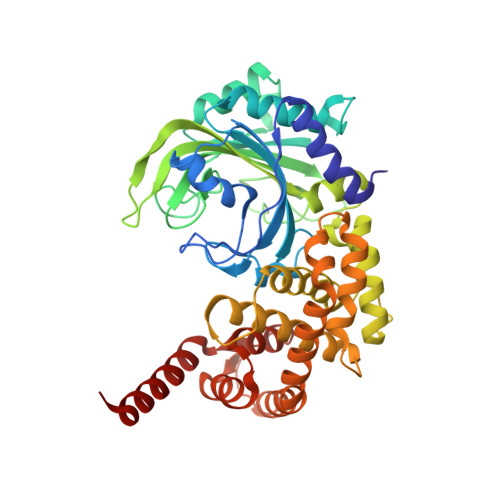
3HXU

Crystal Structure of catalytic fragment of E. coli AlaRS in complex with AlaSA


Experimental Data Snapshot

  • Method: X-RAY DIFFRACTION
  • Resolution: 2.10 Å
  • R-Value Free: 0.191 
  • R-Value Work: 0.154 
  • R-Value Observed: 0.155 

wwPDB Validation   3D Report Full Report


Ligand Structure Quality Assessment 


This is version 1.4 of the entry. See complete history


Literature

Paradox of mistranslation of serine for alanine caused by AlaRS recognition dilemma.

Guo, M.Chong, Y.E.Shapiro, R.Beebe, K.Yang, X.L.Schimmel, P.

(2009) Nature 462: 808-812

  • DOI: https://doi.org/10.1038/nature08612
  • Primary Citation of Related Structures:  
    3HXU, 3HXV, 3HXW, 3HXX, 3HXY, 3HXZ, 3HY0, 3HY1

  • PubMed Abstract: 

    Mistranslation arising from confusion of serine for alanine by alanyl-tRNA synthetases (AlaRSs) has profound functional consequences. Throughout evolution, two editing checkpoints prevent disease-causing mistranslation from confusing glycine or serine for alanine at the active site of AlaRS. In both bacteria and mice, Ser poses a bigger challenge than Gly. One checkpoint is the AlaRS editing centre, and the other is from widely distributed AlaXps-free-standing, genome-encoded editing proteins that clear Ser-tRNA(Ala). The paradox of misincorporating both a smaller (glycine) and a larger (serine) amino acid suggests a deep conflict for nature-designed AlaRS. Here we show the chemical basis for this conflict. Nine crystal structures, together with kinetic and mutational analysis, provided snapshots of adenylate formation for each amino acid. An inherent dilemma is posed by constraints of a structural design that pins down the alpha-amino group of the bound amino acid by using an acidic residue. This design, dating back more than 3 billion years, creates a serendipitous interaction with the serine OH that is difficult to avoid. Apparently because no better architecture for the recognition of alanine could be found, the serine misactivation problem was solved through free-standing AlaXps, which appeared contemporaneously with early AlaRSs. The results reveal unconventional problems and solutions arising from the historical design of the protein synthesis machinery.


  • Organizational Affiliation

    The Skaggs Institute for Chemical Biology and Department of Molecular Biology, The Scripps Research Institute, BCC-379, 10550 North Torrey Pines Road, La Jolla, California 92037, USA.


Macromolecules
Find similar proteins by:  (by identity cutoff)  |  3D Structure
Entity ID: 1
MoleculeChains Sequence LengthOrganismDetailsImage
Alanyl-tRNA synthetase441Escherichia coli K-12Mutation(s): 3 
Gene Names: alaSlovBb2697JW2667
EC: 6.1.1.7 (PDB Primary Data), 6 (UniProt)
UniProt
Find proteins for P00957 (Escherichia coli (strain K12))
Explore P00957 
Go to UniProtKB:  P00957
Entity Groups  
Sequence Clusters30% Identity50% Identity70% Identity90% Identity95% Identity100% Identity
UniProt GroupP00957
Sequence Annotations
Expand
  • Reference Sequence
Small Molecules
Ligands 4 Unique
IDChains Name / Formula / InChI Key2D Diagram3D Interactions
A5A
Query on A5A

Download Ideal Coordinates CCD File 
B [auth A]'5'-O-(N-(L-ALANYL)-SULFAMOYL)ADENOSINE
C13 H19 N7 O7 S
CWWYMWDIYBJVLP-YTMOPEAISA-N
EPE
Query on EPE

Download Ideal Coordinates CCD File 
D [auth A]4-(2-HYDROXYETHYL)-1-PIPERAZINE ETHANESULFONIC ACID
C8 H18 N2 O4 S
JKMHFZQWWAIEOD-UHFFFAOYSA-N
HED
Query on HED

Download Ideal Coordinates CCD File 
C [auth A]2-HYDROXYETHYL DISULFIDE
C4 H10 O2 S2
KYNFOMQIXZUKRK-UHFFFAOYSA-N
BME
Query on BME

Download Ideal Coordinates CCD File 
E [auth A],
F [auth A]
BETA-MERCAPTOETHANOL
C2 H6 O S
DGVVWUTYPXICAM-UHFFFAOYSA-N
Experimental Data & Validation

Experimental Data

  • Method: X-RAY DIFFRACTION
  • Resolution: 2.10 Å
  • R-Value Free: 0.191 
  • R-Value Work: 0.154 
  • R-Value Observed: 0.155 
  • Space Group: P 41 21 2
Unit Cell:
Length ( Å )Angle ( ˚ )
a = 114.754α = 90
b = 114.754β = 90
c = 125.37γ = 90
Software Package:
Software NamePurpose
DENZOdata reduction
SCALEPACKdata scaling
MOLREPphasing
REFMACrefinement
PDB_EXTRACTdata extraction
HKL-2000data collection

Structure Validation

View Full Validation Report


Ligand Structure Quality Assessment 


Entry History 

Deposition Data

Revision History  (Full details and data files)

  • Version 1.0: 2009-12-15
    Type: Initial release
  • Version 1.1: 2011-07-13
    Changes: Version format compliance
  • Version 1.2: 2017-11-01
    Changes: Refinement description
  • Version 1.3: 2021-10-13
    Changes: Database references, Derived calculations
  • Version 1.4: 2024-02-21
    Changes: Data collection