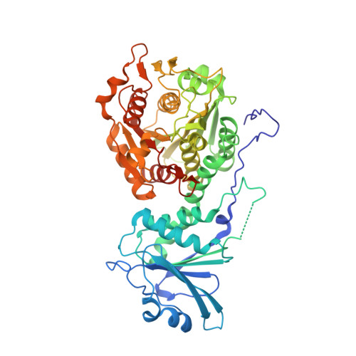
3KQZ

Structure of a protease 2


Experimental Data Snapshot

  • Method: X-RAY DIFFRACTION
  • Resolution: 2.39 Å
  • R-Value Free: 0.242 
  • R-Value Work: 0.176 
  • R-Value Observed: 0.179 

Starting Model: experimental
View more details

wwPDB Validation   3D Report Full Report


This is version 1.3 of the entry. See complete history


Literature

Structure of the Plasmodium falciparum M17 aminopeptidase and significance for the design of drugs targeting the neutral exopeptidases

McGowan, S.Oellig, C.A.Birru, W.A.Caradoc-Davies, T.T.Stack, C.M.Lowther, J.Skinner-Adams, T.Mucha, A.Kafarski, P.Grembecka, J.Trenholme, K.R.Buckle, A.M.Gardiner, D.L.Dalton, J.P.Whisstock, J.C.

(2010) Proc Natl Acad Sci U S A 107: 2449-2454

  • DOI: https://doi.org/10.1073/pnas.0911813107
  • Primary Citation of Related Structures:  
    3KQX, 3KQZ, 3KR4, 3KR5

  • PubMed Abstract: 

    Current therapeutics and prophylactics for malaria are under severe challenge as a result of the rapid emergence of drug-resistant parasites. The human malaria parasite Plasmodium falciparum expresses two neutral aminopeptidases, PfA-M1 and PfA-M17, which function in regulating the intracellular pool of amino acids required for growth and development inside the red blood cell. These enzymes are essential for parasite viability and are validated therapeutic targets. We previously reported the X-ray crystal structure of the monomeric PfA-M1 and proposed a mechanism for substrate entry and free amino acid release from the active site. Here, we present the X-ray crystal structure of the hexameric leucine aminopeptidase, PfA-M17, alone and in complex with two inhibitors with antimalarial activity. The six active sites of the PfA-M17 hexamer are arranged in a disc-like fashion so that they are orientated inwards to form a central catalytic cavity; flexible loops that sit at each of the six entrances to the catalytic cavern function to regulate substrate access. In stark contrast to PfA-M1, PfA-M17 has a narrow and hydrophobic primary specificity pocket which accounts for its highly restricted substrate specificity. We also explicate the essential roles for the metal-binding centers in these enzymes (two in PfA-M17 and one in PfA-M1) in both substrate and drug binding. Our detailed understanding of the PfA-M1 and PfA-M17 active sites now permits a rational approach in the development of a unique class of two-target and/or combination antimalarial therapy.


  • Organizational Affiliation

    Department of Biochemistry and Molecular Biology, Monash University, Clayton Campus, Melbourne, Victoria 3800, Australia. Sheena.McGowan@med.monash.edu.au


Macromolecules
Find similar proteins by:  (by identity cutoff)  |  3D Structure
Entity ID: 1
MoleculeChains Sequence LengthOrganismDetailsImage
M17 leucyl aminopeptidase
A, B, C, D, E
A, B, C, D, E, F, G, H, I, J, K, L
528Plasmodium falciparum 3D7Mutation(s): 3 
EC: 3.4.11.1 (PDB Primary Data), 3.4.13 (UniProt)
UniProt
Find proteins for Q8IL11 (Plasmodium falciparum (isolate 3D7))
Explore Q8IL11 
Go to UniProtKB:  Q8IL11
Entity Groups  
Sequence Clusters30% Identity50% Identity70% Identity90% Identity95% Identity100% Identity
UniProt GroupQ8IL11
Sequence Annotations
Expand
  • Reference Sequence
Small Molecules
Ligands 5 Unique
IDChains Name / Formula / InChI Key2D Diagram3D Interactions
2PE
Query on 2PE

Download Ideal Coordinates CCD File 
BA [auth B],
JC [auth H],
TB [auth F]
NONAETHYLENE GLYCOL
C18 H38 O10
YZUUTMGDONTGTN-UHFFFAOYSA-N
1PE
Query on 1PE

Download Ideal Coordinates CCD File 
AC [auth G]
AD [auth J]
AE [auth L]
BC [auth G]
BD [auth J]
AC [auth G],
AD [auth J],
AE [auth L],
BC [auth G],
BD [auth J],
CA [auth B],
CC [auth G],
CD [auth J],
DC [auth G],
DD [auth J],
EC [auth G],
FB [auth E],
GB [auth E],
HB [auth E],
IB [auth E],
JB [auth E],
KA [auth C],
KB [auth E],
KC [auth H],
LA [auth C],
MA [auth C],
MD [auth K],
ND [auth K],
OD [auth K],
PB [auth F],
PD [auth K],
QB [auth F],
QC [auth I],
QD [auth K],
R [auth A],
RB [auth F],
RC [auth I],
RD [auth K],
S [auth A],
SB [auth F],
SC [auth I],
SD [auth K],
T [auth A],
TA [auth D],
TC [auth I],
U [auth A],
UA [auth D],
VA [auth D],
WA [auth D],
WD [auth L],
XA [auth D],
XD [auth L],
YA [auth D],
YC [auth J],
YD [auth L],
ZB [auth G],
ZC [auth J],
ZD [auth L]
PENTAETHYLENE GLYCOL
C10 H22 O6
JLFNLZLINWHATN-UHFFFAOYSA-N
SO4
Query on SO4

Download Ideal Coordinates CCD File 
AA [auth B]
CB [auth E]
DB [auth E]
EB [auth E]
GA [auth C]
AA [auth B],
CB [auth E],
DB [auth E],
EB [auth E],
GA [auth C],
HA [auth C],
HD [auth K],
IA [auth C],
IC [auth H],
ID [auth K],
JA [auth C],
JD [auth K],
KD [auth K],
LD [auth K],
OB [auth F],
OC [auth I],
P [auth A],
PC [auth I],
Q [auth A],
QA [auth D],
RA [auth D],
SA [auth D],
XB [auth G],
XC [auth J],
Y [auth B],
YB [auth G],
Z [auth B]
SULFATE ION
O4 S
QAOWNCQODCNURD-UHFFFAOYSA-L
ZN
Query on ZN

Download Ideal Coordinates CCD File 
AB [auth E]
BB [auth E]
EA [auth C]
FA [auth C]
FD [auth K]
AB [auth E],
BB [auth E],
EA [auth C],
FA [auth C],
FD [auth K],
GC [auth H],
GD [auth K],
HC [auth H],
MB [auth F],
MC [auth I],
N [auth A],
NB [auth F],
NC [auth I],
O [auth A],
OA [auth D],
PA [auth D],
UD [auth L],
VB [auth G],
VC [auth J],
VD [auth L],
W [auth B],
WB [auth G],
WC [auth J],
X [auth B]
ZINC ION
Zn
PTFCDOFLOPIGGS-UHFFFAOYSA-N
CO3
Query on CO3

Download Ideal Coordinates CCD File 
DA [auth C]
ED [auth K]
FC [auth H]
LB [auth F]
LC [auth I]
DA [auth C],
ED [auth K],
FC [auth H],
LB [auth F],
LC [auth I],
M [auth A],
NA [auth D],
TD [auth L],
UB [auth G],
UC [auth J],
V [auth B],
ZA [auth E]
CARBONATE ION
C O3
BVKZGUZCCUSVTD-UHFFFAOYSA-L
Experimental Data & Validation

Experimental Data

  • Method: X-RAY DIFFRACTION
  • Resolution: 2.39 Å
  • R-Value Free: 0.242 
  • R-Value Work: 0.176 
  • R-Value Observed: 0.179 
  • Space Group: P 21 21 21
Unit Cell:
Length ( Å )Angle ( ˚ )
a = 171.698α = 90
b = 173.728β = 90
c = 220.278γ = 90
Software Package:
Software NamePurpose
Blu-Icedata collection
MOLREPphasing
REFMACrefinement
MOSFLMdata reduction
SCALAdata scaling

Structure Validation

View Full Validation Report


Entry History 

Deposition Data

Revision History  (Full details and data files)

  • Version 1.0: 2010-02-02
    Type: Initial release
  • Version 1.1: 2011-07-13
    Changes: Version format compliance
  • Version 1.2: 2021-11-10
    Changes: Database references, Derived calculations
  • Version 1.3: 2023-11-01
    Changes: Data collection, Refinement description