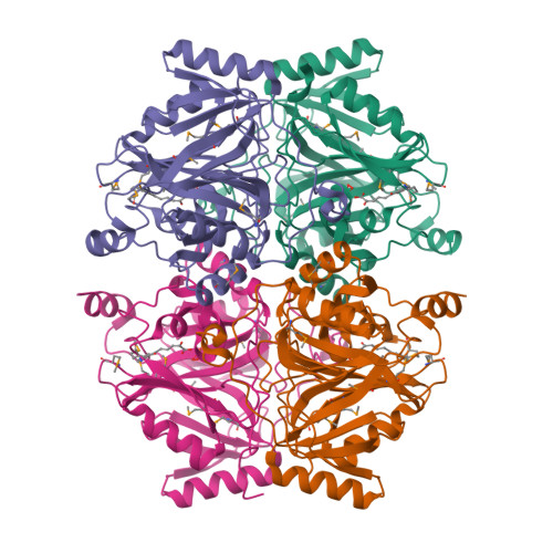
Crystal Structure of 2,3-Dihydroxy Biphenyl dioxygenase from Rhodococcus sp. (strain RHA1)
Syed Ibrahim, B., Kumaran, D., Burley, S.K., Swaminathan, S.To be published.
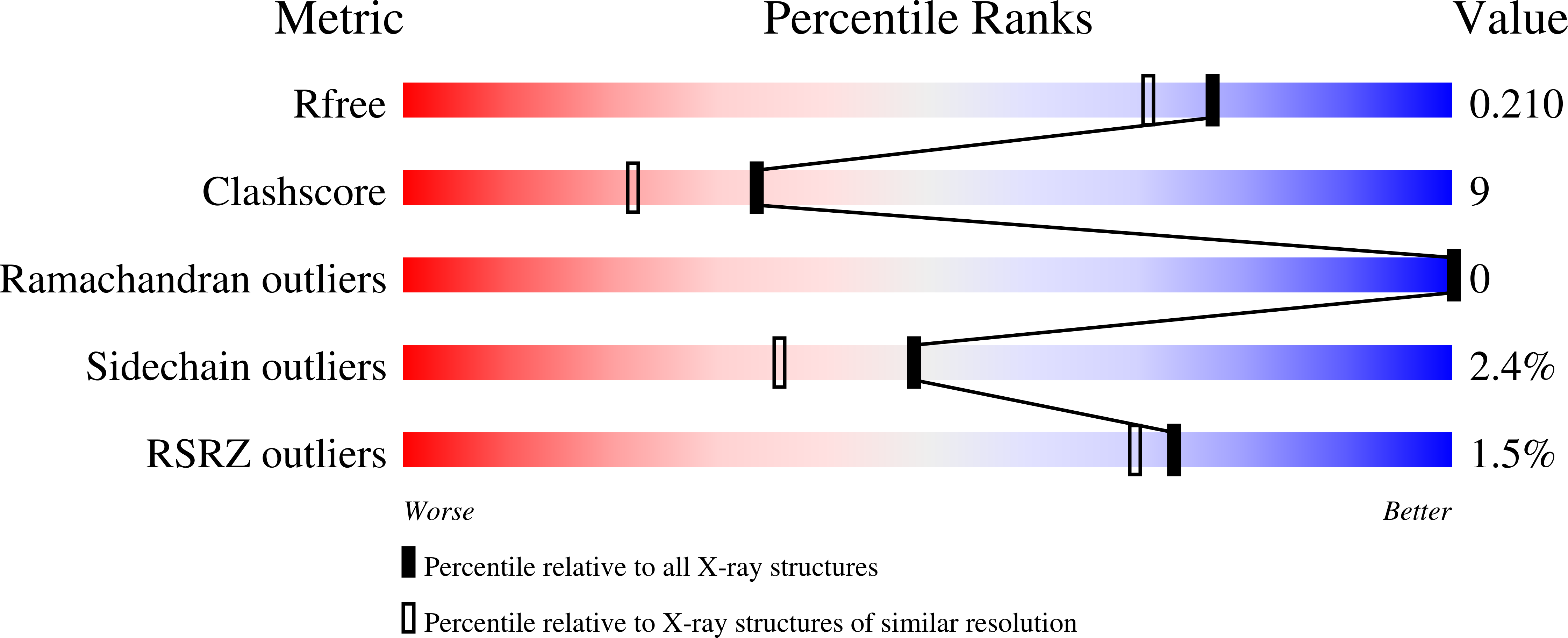
Experimental Data Snapshot
Entity ID: 1 | |||||
|---|---|---|---|---|---|
| Molecule | Chains | Sequence Length | Organism | Details | Image |
| Catechol 2,3-dioxygenase | 339 | Rhodococcus jostii RHA1 | Mutation(s): 0 EC: 1.13.11.2 | 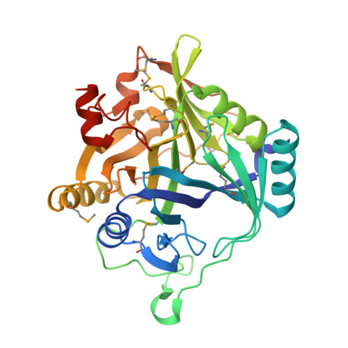 | |
UniProt | |||||
Find proteins for Q0S9X1 (Rhodococcus jostii (strain RHA1)) Explore Q0S9X1 Go to UniProtKB: Q0S9X1 | |||||
Entity Groups | |||||
| Sequence Clusters | 30% Identity50% Identity70% Identity90% Identity95% Identity100% Identity | ||||
| UniProt Group | Q0S9X1 | ||||
Sequence AnnotationsExpand | |||||
| |||||
| Ligands 3 Unique | |||||
|---|---|---|---|---|---|
| ID | Chains | Name / Formula / InChI Key | 2D Diagram | 3D Interactions | |
| HPX Query on HPX | G [auth A], J [auth B], M [auth C], P [auth D] | (2Z,4E)-2-HYDROXY-6-OXO-6-PHENYLHEXA-2,4-DIENOIC ACID C12 H10 O4 RDRDHXDYMGUCKE-KXBBGWRGSA-N |  | ||
| FE Query on FE | E [auth A], H [auth B], K [auth C], N [auth D] | FE (III) ION Fe VTLYFUHAOXGGBS-UHFFFAOYSA-N |  | ||
| PEO Query on PEO | F [auth A], I [auth B], L [auth C], O [auth D] | HYDROGEN PEROXIDE H2 O2 MHAJPDPJQMAIIY-UHFFFAOYSA-N |  | ||
| Modified Residues 1 Unique | |||||
|---|---|---|---|---|---|
| ID | Chains | Type | Formula | 2D Diagram | Parent |
| MSE Query on MSE | A, B, C, D | L-PEPTIDE LINKING | C5 H11 N O2 Se |  | MET |
| Length ( Å ) | Angle ( ˚ ) |
|---|---|
| a = 74.923 | α = 90 |
| b = 76.189 | β = 90 |
| c = 251.373 | γ = 90 |
| Software Name | Purpose |
|---|---|
| CBASS | data collection |
| SHELX | model building |
| SHARP | phasing |
| CNS | refinement |
| HKL-2000 | data reduction |
| SCALEPACK | data scaling |
| SHELX | phasing |
RCSB PDB is hosted by

RCSB PDB is a member of the