3NVX | pdb_00003nvx

Molecular mechanism of guidance cue recognition


Experimental Data Snapshot

  • Method: X-RAY DIFFRACTION
  • Resolution: 2.00 Å
  • R-Value Free: 
    0.225 (Depositor), 0.220 (DCC) 
  • R-Value Work: 
    0.176 (Depositor), 0.172 (DCC) 
  • R-Value Observed: 
    0.178 (Depositor) 

wwPDB Validation   3D Report Full Report


Ligand Structure Quality Assessment 

Created with Raphaël 2.3.0Worse 01 BetterLigand structure goodness of fit to experimental dataBest fitted NAGClick on this verticalbar to view details

This is version 1.3 of the entry. See complete history


Literature

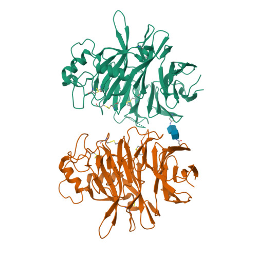
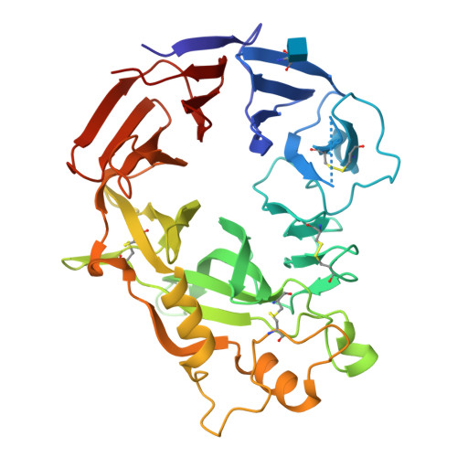
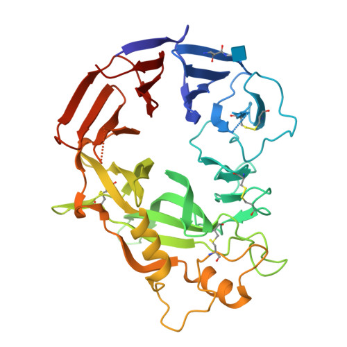
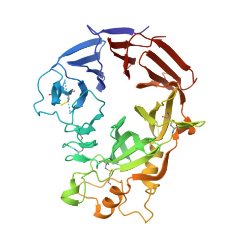
Structural Basis of Semaphorin-Plexin Recognition and Viral Mimicry from Sema7A and A39R Complexes with PlexinC1.

Liu, H.Juo, Z.S.Shim, A.H.Focia, P.J.Chen, X.Garcia, K.C.He, X.

(2010) Cell 142: 749-761

  • DOI: https://doi.org/10.1016/j.cell.2010.07.040
  • Primary Citation of Related Structures:  
    3NVN, 3NVQ, 3NVX

  • PubMed Abstract: 

    Repulsive signaling by Semaphorins and Plexins is crucial for the development and homeostasis of the nervous, immune, and cardiovascular systems. Sema7A acts as both an immune and a neural Semaphorin through PlexinC1, and A39R is a Sema7A mimic secreted by smallpox virus. We report the structures of Sema7A and A39R complexed with the Semaphorin-binding module of PlexinC1. Both structures show two PlexinC1 molecules symmetrically bridged by Semaphorin dimers, in which the Semaphorin and PlexinC1 beta propellers interact in an edge-on, orthogonal orientation. Both binding interfaces are dominated by the insertion of the Semaphorin's 4c-4d loop into a deep groove in blade 3 of the PlexinC1 propeller. A39R appears to achieve Sema7A mimicry by preserving key Plexin-binding determinants seen in the mammalian Sema7A complex that have evolved to achieve higher affinity binding to the host-derived PlexinC1. The complex structures support a conserved Semaphorin-Plexin recognition mode and suggest that Plexins are activated by dimerization.


  • Organizational Affiliation

    Department of Molecular Pharmacology and Biological Chemistry, Northwestern University Feinberg School of Medicine, Chicago, IL 60611, USA.


Macromolecules
Find similar proteins by:  (by identity cutoff)  |  3D Structure
Entity ID: 1
MoleculeChains Sequence LengthOrganismDetailsImage
Protein A39
A, B
383Vaccinia virus CopenhagenMutation(s): 0 
Gene Names: A39R
UniProt
Find proteins for P21062 (Vaccinia virus (strain Copenhagen))
Explore P21062 
Go to UniProtKB:  P21062
Entity Groups  
Sequence Clusters30% Identity50% Identity70% Identity90% Identity95% Identity100% Identity
UniProt GroupP21062
Glycosylation
Glycosylation Sites: 1
Sequence Annotations
Expand
  • Reference Sequence
Experimental Data & Validation

Experimental Data

  • Method: X-RAY DIFFRACTION
  • Resolution: 2.00 Å
  • R-Value Free:  0.225 (Depositor), 0.220 (DCC) 
  • R-Value Work:  0.176 (Depositor), 0.172 (DCC) 
  • R-Value Observed: 0.178 (Depositor) 
Space Group: C 2 2 21
Unit Cell:
Length ( Å )Angle ( ˚ )
a = 51.088α = 90
b = 171.006β = 90
c = 211.175γ = 90
Software Package:
Software NamePurpose
HKL-2000data collection
SOLVEphasing
PHENIXrefinement
MOSFLMdata reduction
SCALAdata scaling

Structure Validation

View Full Validation Report


Ligand Structure Quality Assessment 

Created with Raphaël 2.3.0Worse 01 BetterLigand structure goodness of fit to experimental dataBest fitted NAGClick on this verticalbar to view details

Entry History 

Deposition Data

Revision History  (Full details and data files)

  • Version 1.0: 2010-09-15
    Type: Initial release
  • Version 1.1: 2011-07-13
    Changes: Source and taxonomy, Version format compliance
  • Version 1.2: 2020-07-29
    Type: Remediation
    Reason: Carbohydrate remediation
    Changes: Data collection, Database references, Derived calculations, Structure summary
  • Version 1.3: 2024-10-30
    Changes: Data collection, Database references, Structure summary