3OIG

Crystal Structure of Enoyl-ACP Reductases I (FabI) from B. subtilis (complex with NAD and INH)


Experimental Data Snapshot

  • Method: X-RAY DIFFRACTION
  • Resolution: 1.25 Å
  • R-Value Free: 
    0.185 (Depositor), 0.180 (DCC) 
  • R-Value Work: 
    0.167 (Depositor), 0.160 (DCC) 
  • R-Value Observed: 
    0.167 (Depositor) 

Starting Model: experimental
View more details

wwPDB Validation   3D Report Full Report


Ligand Structure Quality Assessment 

Created with Raphaël 2.3.0Worse 01 BetterLigand structure goodness of fit to experimental dataBest fitted NADClick on this verticalbar to view detailsBest fitted IMJClick on this verticalbar to view details

This is version 1.2 of the entry. See complete history


Literature

Crystal Structures of Enoyl-ACP Reductases I (FabI) and III (FabL) from B. subtilis

Kim, K.-H.Ha, B.H.Kim, S.J.Hong, S.K.Hwang, K.Y.Kim, E.E.

(2011) J Mol Biol 406: 403-415

  • DOI: https://doi.org/10.1016/j.jmb.2010.12.003
  • Primary Citation of Related Structures:  
    3OIC, 3OID, 3OIF, 3OIG

  • PubMed Abstract: 

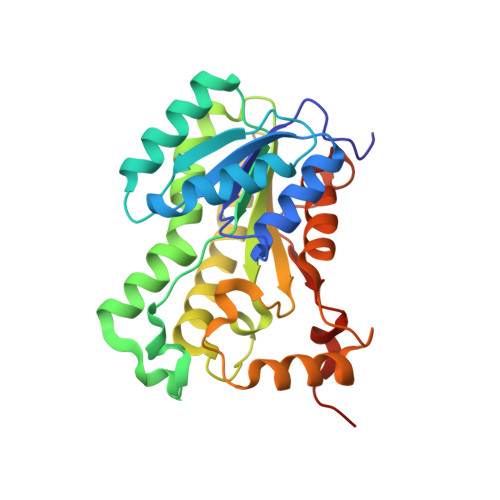
    Enoyl-[acyl carrier protein] (ACP) reductase (ENR) is a key enzyme in type II fatty acid synthesis that catalyzes the last step in each elongation cycle. Therefore, it has been considered as a target for antibiotics. However, recent studies indicate that some pathogens have more than one ENR; in particular, Bacillus subtilis has two ENRs, FabI and FabL. The crystal structures of the ternary complexes of BsFaBI and BsFabL are found as a homotetramer showing the same overall structure despite a sequence identity of only 24%. The positions of the catalytic dyad of Tyr-(Xaa)(6)-Lys in FabL are almost identical to that of FabI, but a detailed structural analysis shows that FabL shares more structural similarities with FabG and other members of the SDR (short-chain alcohol dehydrogenase/reductase) family. The apo FabL structure shows significantly different conformations at the cofactor and the substrate-binding regions, and this resulted in a totally different tetrameric arrangement reflecting the flexibility of these regions in the absence of the cofactor and substrate/inhibitor.


  • Organizational Affiliation

    Life Sciences Division, Korea Institute of Science and Technology, 39-1 Hawolkok-dong, Sungbuk-gu, Seoul 136-791, South Korea.


Macromolecules
Find similar proteins by:  (by identity cutoff)  |  3D Structure
Entity ID: 1
MoleculeChains Sequence LengthOrganismDetailsImage
Enoyl-[acyl-carrier-protein] reductase [NADH]266Bacillus subtilisMutation(s): 0 
Gene Names: FabI
EC: 1.3.1.9
UniProt
Find proteins for P54616 (Bacillus subtilis (strain 168))
Explore P54616 
Go to UniProtKB:  P54616
Entity Groups  
Sequence Clusters30% Identity50% Identity70% Identity90% Identity95% Identity100% Identity
UniProt GroupP54616
Sequence Annotations
Expand
  • Reference Sequence
Small Molecules
Ligands 2 Unique
IDChains Name / Formula / InChI Key2D Diagram3D Interactions
NAD
Query on NAD

Download Ideal Coordinates CCD File 
B [auth A]NICOTINAMIDE-ADENINE-DINUCLEOTIDE
C21 H27 N7 O14 P2
BAWFJGJZGIEFAR-NNYOXOHSSA-N
IMJ
Query on IMJ

Download Ideal Coordinates CCD File 
C [auth A](2E)-N-[(1,2-dimethyl-1H-indol-3-yl)methyl]-N-methyl-3-(7-oxo-5,6,7,8-tetrahydro-1,8-naphthyridin-3-yl)prop-2-enamide
C23 H24 N4 O2
PVNPCRMKZHRPEV-DHZHZOJOSA-N
Experimental Data & Validation

Experimental Data

  • Method: X-RAY DIFFRACTION
  • Resolution: 1.25 Å
  • R-Value Free:  0.185 (Depositor), 0.180 (DCC) 
  • R-Value Work:  0.167 (Depositor), 0.160 (DCC) 
  • R-Value Observed: 0.167 (Depositor) 
Space Group: I 2 2 2
Unit Cell:
Length ( Å )Angle ( ˚ )
a = 69.126α = 90
b = 80.004β = 90
c = 88.077γ = 90
Software Package:
Software NamePurpose
PROTEUM PLUSdata collection
CNSrefinement
REFMACrefinement
HKL-2000data reduction
HKL-2000data scaling
CNSphasing

Structure Validation

View Full Validation Report


Ligand Structure Quality Assessment 

Created with Raphaël 2.3.0Worse 01 BetterLigand structure goodness of fit to experimental dataBest fitted NADClick on this verticalbar to view detailsBest fitted IMJClick on this verticalbar to view details

Entry History 

Deposition Data

Revision History  (Full details and data files)

  • Version 1.0: 2011-01-05
    Type: Initial release
  • Version 1.1: 2011-07-13
    Changes: Version format compliance
  • Version 1.2: 2023-11-01
    Changes: Data collection, Database references, Derived calculations, Refinement description