3QUZ

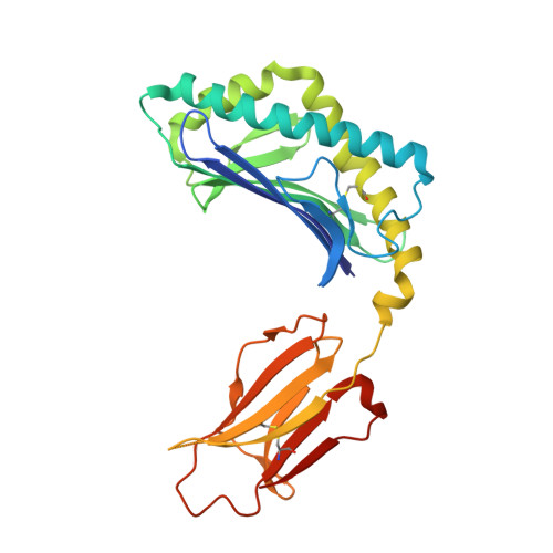
Structure of the mouse CD1d-NU-alpha-GalCer-iNKT TCR complex


Experimental Data Snapshot

  • Method: X-RAY DIFFRACTION
  • Resolution: 2.30 Å
  • R-Value Free: 0.233 
  • R-Value Work: 0.192 
  • R-Value Observed: 0.194 

Starting Models: experimental
View more details

wwPDB Validation   3D Report Full Report


Ligand Structure Quality Assessment 


This is version 2.2 of the entry. See complete history


Literature

Galactose-modified iNKT cell agonists stabilized by an induced fit of CD1d prevent tumour metastasis.

Aspeslagh, S.Li, Y.Yu, E.D.Pauwels, N.Trappeniers, M.Girardi, E.Decruy, T.Van Beneden, K.Venken, K.Drennan, M.Leybaert, L.Wang, J.Franck, R.W.Van Calenbergh, S.Zajonc, D.M.Elewaut, D.

(2011) EMBO J 30: 2294-2305

  • DOI: https://doi.org/10.1038/emboj.2011.145
  • Primary Citation of Related Structures:  
    3QUX, 3QUY, 3QUZ

  • PubMed Abstract: 

    Invariant natural killer T (iNKT) cells are known to have marked immunomodulatory capacity due to their ability to produce copious amounts of effector cytokines. Here, we report the structure and function of a novel class of aromatic α-galactosylceramide structurally related glycolipids with marked Th1 bias in both mice and men, leading to superior tumour protection in vivo. The strength of the Th1 response correlates well with enhanced lipid binding to CD1d as a result of an induced fit mechanism that binds the aromatic substitution as a third anchor, in addition to the two lipid chains. This induced fit is in contrast to another Th1-biasing glycolipid, α-C-GalCer, whose CD1d binding follows a conventional key-lock principle. These findings highlight the previously unexploited flexibility of CD1d in accommodating galactose-modified glycolipids and broaden the range of glycolipids that can stimulate iNKT cells. We speculate that glycolipids can be designed that induce a similar fit, thereby leading to superior and more sustained iNKT cell responses in vivo.


  • Organizational Affiliation

    Department of Rheumatology, Faculty of Medicine and Health Sciences, Laboratory for Molecular Immunology and Inflammation, Ghent University, Ghent, Belgium.


Macromolecules
Find similar proteins by:  (by identity cutoff)  |  3D Structure
Entity ID: 1
MoleculeChains Sequence LengthOrganismDetailsImage
Antigen-presenting glycoprotein CD1d1285Mus musculusMutation(s): 0 
Gene Names: Cd1.1CD1dCd1d1
UniProt & NIH Common Fund Data Resources
Find proteins for P11609 (Mus musculus)
Explore P11609 
Go to UniProtKB:  P11609
IMPC:  MGI:107674
Entity Groups  
Sequence Clusters30% Identity50% Identity70% Identity90% Identity95% Identity100% Identity
UniProt GroupP11609
Glycosylation
Glycosylation Sites: 3Go to GlyGen: P11609-1
Sequence Annotations
Expand
  • Reference Sequence
Find similar proteins by:  (by identity cutoff)  |  3D Structure
Entity ID: 2
MoleculeChains Sequence LengthOrganismDetailsImage
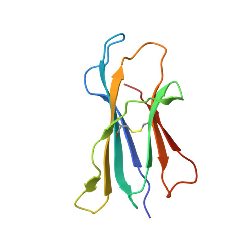
Beta-2 microglobulin99Mus musculusMutation(s): 0 
Gene Names: B2mbeta-2-microglobulinmCG_11606RP23-34E24.5-001
UniProt & NIH Common Fund Data Resources
Find proteins for P01887 (Mus musculus)
Explore P01887 
Go to UniProtKB:  P01887
IMPC:  MGI:88127
Entity Groups  
Sequence Clusters30% Identity50% Identity70% Identity90% Identity95% Identity100% Identity
UniProt GroupP01887
Sequence Annotations
Expand
  • Reference Sequence
Find similar proteins by:  (by identity cutoff)  |  3D Structure
Entity ID: 3
MoleculeChains Sequence LengthOrganismDetailsImage
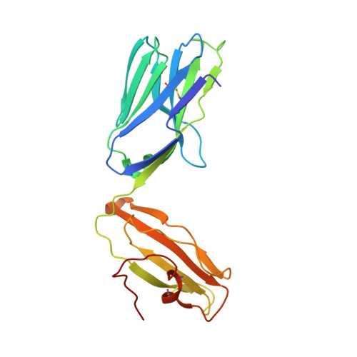
Valpha14 (mouse variable domain, human constant domain)209Mus musculusMutation(s): 0 
Entity Groups  
Sequence Clusters30% Identity50% Identity70% Identity90% Identity95% Identity100% Identity
Sequence Annotations
Expand
  • Reference Sequence
Find similar proteins by:  (by identity cutoff)  |  3D Structure
Entity ID: 4
MoleculeChains Sequence LengthOrganismDetailsImage
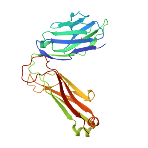
Vbeta8.2 (mouse variable domain, human constant domain)241Mus musculusMutation(s): 0 
Entity Groups  
Sequence Clusters30% Identity50% Identity70% Identity90% Identity95% Identity100% Identity
Sequence Annotations
Expand
  • Reference Sequence
Oligosaccharides

Help

Entity ID: 5
MoleculeChains Length2D Diagram Glycosylation3D Interactions
2-acetamido-2-deoxy-beta-D-glucopyranose-(1-4)-[alpha-L-fucopyranose-(1-6)]2-acetamido-2-deoxy-beta-D-glucopyranose
E
3N-Glycosylation
Glycosylation Resources
GlyTouCan:  G21290RB
GlyCosmos:  G21290RB
GlyGen:  G21290RB
Small Molecules
Ligands 2 Unique
IDChains Name / Formula / InChI Key2D Diagram3D Interactions
QUV
Query on QUV

Download Ideal Coordinates CCD File 
H [auth A]N-[(2S,3S,4R)-1-({6-deoxy-6-[(naphthalen-1-ylcarbamoyl)amino]-alpha-D-galactopyranosyl}oxy)-3,4-dihydroxyoctadecan-2-yl]hexacosanamide
C61 H107 N3 O9
LYMLSPSPEXPNPQ-DBQLOCGYSA-N
NAG
Query on NAG

Download Ideal Coordinates CCD File 
F [auth A],
G [auth A]
2-acetamido-2-deoxy-beta-D-glucopyranose
C8 H15 N O6
OVRNDRQMDRJTHS-FMDGEEDCSA-N
Binding Affinity Annotations 
IDSourceBinding Affinity
QUV PDBBind:  3QUZ Kd: 39.6 (nM) from 1 assay(s)
Experimental Data & Validation

Experimental Data

  • Method: X-RAY DIFFRACTION
  • Resolution: 2.30 Å
  • R-Value Free: 0.233 
  • R-Value Work: 0.192 
  • R-Value Observed: 0.194 
  • Space Group: C 2 2 21
Unit Cell:
Length ( Å )Angle ( ˚ )
a = 79.292α = 90
b = 190.981β = 90
c = 151.318γ = 90
Software Package:
Software NamePurpose
HKL-2000data collection
MOLREPphasing
REFMACrefinement
HKL-2000data reduction
HKL-2000data scaling

Structure Validation

View Full Validation Report


Ligand Structure Quality Assessment 


Entry History 

Deposition Data

Revision History  (Full details and data files)

  • Version 1.0: 2011-06-29
    Type: Initial release
  • Version 1.1: 2011-07-13
    Changes: Version format compliance
  • Version 2.0: 2020-07-29
    Type: Remediation
    Reason: Carbohydrate remediation
    Changes: Atomic model, Data collection, Database references, Derived calculations, Structure summary
  • Version 2.1: 2023-09-13
    Changes: Data collection, Database references, Refinement description, Structure summary
  • Version 2.2: 2024-10-16
    Changes: Structure summary