3R1K

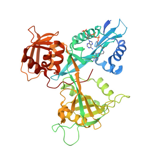
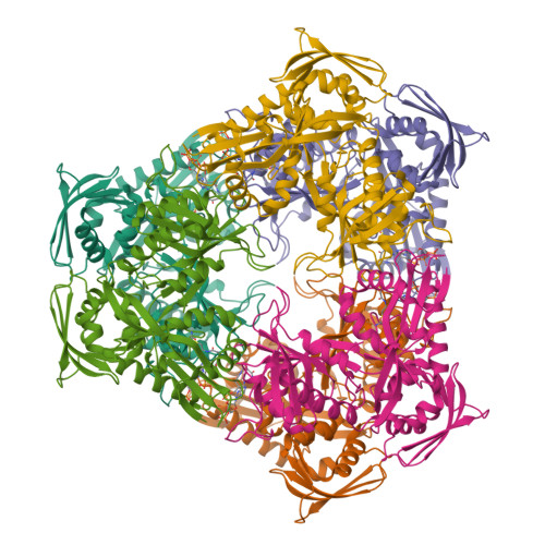
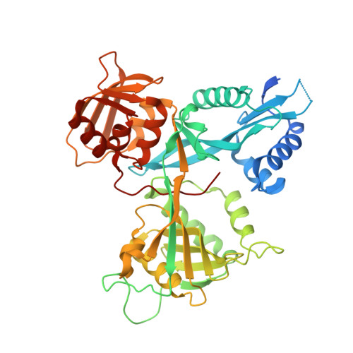
Crystal structure of acetyltransferase Eis from Mycobacterium tuberculosis H37Rv in complex with CoA and an acetamide moiety


Experimental Data Snapshot

  • Method: X-RAY DIFFRACTION
  • Resolution: 1.95 Å
  • R-Value Free: 0.224 
  • R-Value Work: 0.195 
  • R-Value Observed: 0.196 

wwPDB Validation   3D Report Full Report


Ligand Structure Quality Assessment 


This is version 1.2 of the entry. See complete history


Literature

Unusual regioversatility of acetyltransferase Eis, a cause of drug resistance in XDR-TB.

Chen, W.Biswas, T.Porter, V.R.Tsodikov, O.V.Garneau-Tsodikova, S.

(2011) Proc Natl Acad Sci U S A 108: 9804-9808

  • DOI: https://doi.org/10.1073/pnas.1105379108
  • Primary Citation of Related Structures:  
    3R1K

  • PubMed Abstract: 

    The emergence of multidrug-resistant and extensively drug-resistant (XDR) tuberculosis (TB) is a serious global threat. Aminoglycoside antibiotics are used as a last resort to treat XDR-TB. Resistance to the aminoglycoside kanamycin is a hallmark of XDR-TB. Here, we reveal the function and structure of the mycobacterial protein Eis responsible for resistance to kanamycin in a significant fraction of kanamycin-resistant Mycobacterium tuberculosis clinical isolates. We demonstrate that Eis has an unprecedented ability to acetylate multiple amines of many aminoglycosides. Structural and mutagenesis studies of Eis indicate that its acetylation mechanism is enabled by a complex tripartite fold that includes two general control non-derepressible 5 (GCN5)-related N-acetyltransferase regions. An intricate negatively charged substrate-binding pocket of Eis is a potential target of new antitubercular drugs expected to overcome aminoglycoside resistance.


  • Organizational Affiliation

    Life Sciences Institute, University of Michigan, Ann Arbor, MI 48109, USA.


Macromolecules
Find similar proteins by:  (by identity cutoff)  |  3D Structure
Entity ID: 1
MoleculeChains Sequence LengthOrganismDetailsImage
Enhanced intracellular survival protein428Mycobacterium tuberculosisMutation(s): 0 
Gene Names: eisRv2416cMT2489MTCY253.04
EC: 2.3.1
UniProt
Find proteins for P9WFK7 (Mycobacterium tuberculosis (strain ATCC 25618 / H37Rv))
Explore P9WFK7 
Go to UniProtKB:  P9WFK7
Entity Groups  
Sequence Clusters30% Identity50% Identity70% Identity90% Identity95% Identity100% Identity
UniProt GroupP9WFK7
Sequence Annotations
Expand
  • Reference Sequence
Experimental Data & Validation

Experimental Data

  • Method: X-RAY DIFFRACTION
  • Resolution: 1.95 Å
  • R-Value Free: 0.224 
  • R-Value Work: 0.195 
  • R-Value Observed: 0.196 
  • Space Group: H 3 2
Unit Cell:
Length ( Å )Angle ( ˚ )
a = 174.935α = 90
b = 174.935β = 90
c = 124.686γ = 120
Software Package:
Software NamePurpose
HKL-2000data collection
PHASERphasing
REFMACrefinement
HKL-2000data reduction
HKL-2000data scaling

Structure Validation

View Full Validation Report


Ligand Structure Quality Assessment 


Entry History 

Deposition Data

Revision History  (Full details and data files)

  • Version 1.0: 2011-06-01
    Type: Initial release
  • Version 1.1: 2011-07-13
    Changes: Version format compliance
  • Version 1.2: 2024-02-21
    Changes: Data collection, Database references, Derived calculations