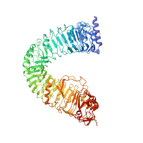
3RJ0

Plant steroid receptor BRI1 ectodomain in complex with brassinolide


Experimental Data Snapshot

  • Method: X-RAY DIFFRACTION
  • Resolution: 2.54 Å
  • R-Value Free: 0.241 
  • R-Value Work: 0.184 
  • R-Value Observed: 0.187 

wwPDB Validation   3D Report Full Report


Ligand Structure Quality Assessment 


This is version 2.0 of the entry. See complete history


Literature

Structural basis of steroid hormone perception by the receptor kinase BRI1.

Hothorn, M.Belkhadir, Y.Dreux, M.Dabi, T.Noel, J.P.Wilson, I.A.Chory, J.

(2011) Nature 474: 467-471

  • DOI: https://doi.org/10.1038/nature10153
  • Primary Citation of Related Structures:  
    3RIZ, 3RJ0

  • PubMed Abstract: 

    Polyhydroxylated steroids are regulators of body shape and size in higher organisms. In metazoans, intracellular receptors recognize these molecules. Plants, however, perceive steroids at membranes, using the membrane-integral receptor kinase BRASSINOSTEROID INSENSITIVE 1 (BRI1). Here we report the structure of the Arabidopsis thaliana BRI1 ligand-binding domain, determined by X-ray diffraction at 2.5 Å resolution. We find a superhelix of 25 twisted leucine-rich repeats (LRRs), an architecture that is strikingly different from the assembly of LRRs in animal Toll-like receptors. A 70-amino-acid island domain between LRRs 21 and 22 folds back into the interior of the superhelix to create a surface pocket for binding the plant hormone brassinolide. Known loss- and gain-of-function mutations map closely to the hormone-binding site. We propose that steroid binding to BRI1 generates a docking platform for a co-receptor that is required for receptor activation. Our findings provide insight into the activation mechanism of this highly expanded family of plant receptors that have essential roles in hormone, developmental and innate immunity signalling.


  • Organizational Affiliation

    Plant Biology Laboratory, The Salk Institute for Biological Studies, 10010 North Torrey Pines Road, La Jolla, California 92037, USA.


Macromolecules
Find similar proteins by:  (by identity cutoff)  |  3D Structure
Entity ID: 1
MoleculeChains Sequence LengthOrganismDetailsImage
Protein BRASSINOSTEROID INSENSITIVE 1772Arabidopsis thalianaMutation(s): 0 
Gene Names: At4g39400BRI1F23K16.30
EC: 2.7.10.1 (PDB Primary Data), 2.7.11.1 (PDB Primary Data)
UniProt
Find proteins for O22476 (Arabidopsis thaliana)
Explore O22476 
Go to UniProtKB:  O22476
Entity Groups  
Sequence Clusters30% Identity50% Identity70% Identity90% Identity95% Identity100% Identity
UniProt GroupO22476
Glycosylation
Glycosylation Sites: 6
Sequence Annotations
Expand
  • Reference Sequence
Oligosaccharides

Help

Entity ID: 2
MoleculeChains Length2D Diagram Glycosylation3D Interactions
alpha-D-mannopyranose-(1-3)-beta-D-mannopyranose-(1-4)-2-acetamido-2-deoxy-beta-D-glucopyranose-(1-4)-2-acetamido-2-deoxy-beta-D-glucopyranose
B, C, D
4N-Glycosylation
Glycosylation Resources
GlyTouCan:  G81315DD
GlyCosmos:  G81315DD
GlyGen:  G81315DD
Experimental Data & Validation

Experimental Data

  • Method: X-RAY DIFFRACTION
  • Resolution: 2.54 Å
  • R-Value Free: 0.241 
  • R-Value Work: 0.184 
  • R-Value Observed: 0.187 
  • Space Group: C 1 2 1
Unit Cell:
Length ( Å )Angle ( ˚ )
a = 175.17α = 90
b = 67.236β = 121.42
c = 119.246γ = 90
Software Package:
Software NamePurpose
StructureStudiodata collection
SHARPphasing
PHENIXrefinement
XDSdata reduction
XSCALEdata scaling

Structure Validation

View Full Validation Report


Ligand Structure Quality Assessment 


Entry History 

Deposition Data

  • Released Date: 2011-06-22 
  • Deposition Author(s): Hothorn, M.

Revision History  (Full details and data files)

  • Version 1.0: 2011-06-22
    Type: Initial release
  • Version 1.1: 2011-07-13
    Changes: Version format compliance
  • Version 2.0: 2020-07-29
    Type: Remediation
    Reason: Carbohydrate remediation
    Changes: Atomic model, Data collection, Database references, Derived calculations, Structure summary