3SYC

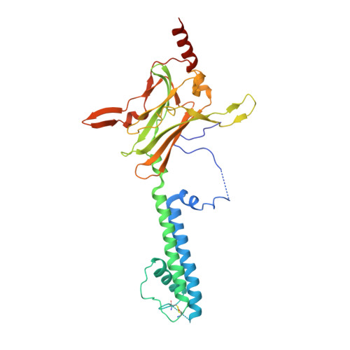
Crystal structure of the G protein-gated inward rectifier K+ channel GIRK2 (Kir3.2) D228N mutant


Experimental Data Snapshot

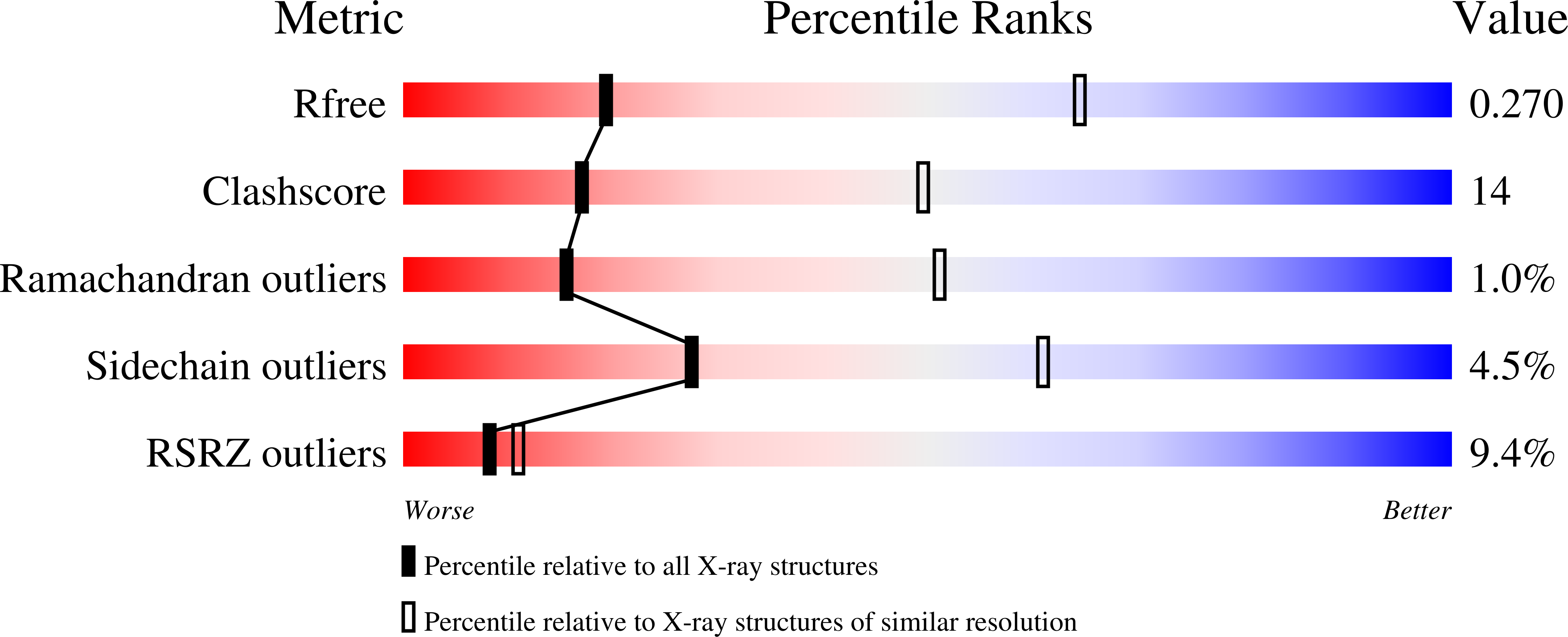
  • Method: X-RAY DIFFRACTION
  • Resolution: 3.41 Å
  • R-Value Free: 0.271 
  • R-Value Work: 0.251 
  • R-Value Observed: 0.252 

Starting Model: experimental
View more details

wwPDB Validation   3D Report Full Report


This is version 1.2 of the entry. See complete history


Literature

Crystal Structure of the Mammalian GIRK2 K(+) Channel and Gating Regulation by G Proteins, PIP(2), and Sodium.

Whorton, M.R.Mackinnon, R.

(2011) Cell 147: 199-208

  • DOI: https://doi.org/10.1016/j.cell.2011.07.046
  • Primary Citation of Related Structures:  
    3SYA, 3SYC, 3SYO, 3SYP, 3SYQ

  • PubMed Abstract: 

    G protein-gated K(+) channels (Kir3.1-Kir3.4) control electrical excitability in many different cells. Among their functions relevant to human physiology and disease, they regulate the heart rate and govern a wide range of neuronal activities. Here, we present the first crystal structures of a G protein-gated K(+) channel. By comparing the wild-type structure to that of a constitutively active mutant, we identify a global conformational change through which G proteins could open a G loop gate in the cytoplasmic domain. The structures of both channels in the absence and presence of PIP(2) suggest that G proteins open only the G loop gate in the absence of PIP(2), but in the presence of PIP(2) the G loop gate and a second inner helix gate become coupled, so that both gates open. We also identify a strategically located Na(+) ion-binding site, which would allow intracellular Na(+) to modulate GIRK channel activity. These data provide a structural basis for understanding multiligand regulation of GIRK channel gating.


  • Organizational Affiliation

    Laboratory of Molecular Neurobiology and Biophysics, Rockefeller University, New York, NY 10065, USA.


Macromolecules
Find similar proteins by:  (by identity cutoff)  |  3D Structure
Entity ID: 1
MoleculeChains Sequence LengthOrganismDetailsImage
G protein-activated inward rectifier potassium channel 2340Mus musculusMutation(s): 1 
Gene Names: Girk2Kcnj6Kcnj7W
Membrane Entity: Yes 
UniProt & NIH Common Fund Data Resources
Find proteins for P48542 (Mus musculus)
Explore P48542 
Go to UniProtKB:  P48542
IMPC:  MGI:104781
Entity Groups  
Sequence Clusters30% Identity50% Identity70% Identity90% Identity95% Identity100% Identity
UniProt GroupP48542
Sequence Annotations
Expand
  • Reference Sequence
Experimental Data & Validation

Experimental Data

  • Method: X-RAY DIFFRACTION
  • Resolution: 3.41 Å
  • R-Value Free: 0.271 
  • R-Value Work: 0.251 
  • R-Value Observed: 0.252 
  • Space Group: P 4 21 2
Unit Cell:
Length ( Å )Angle ( ˚ )
a = 86.488α = 90
b = 86.488β = 90
c = 179.627γ = 90
Software Package:
Software NamePurpose
SCALEPACKdata scaling
REFMACrefinement
PDB_EXTRACTdata extraction
CBASSdata collection
DENZOdata reduction
MOLREPphasing

Structure Validation

View Full Validation Report


Entry History 

Deposition Data

Revision History  (Full details and data files)

  • Version 1.0: 2011-10-12
    Type: Initial release
  • Version 1.1: 2023-09-13
    Changes: Data collection, Database references, Derived calculations, Refinement description
  • Version 1.2: 2024-10-30
    Changes: Structure summary