3TDL

Structure of human serum albumin in complex with DAUDA


Experimental Data Snapshot

  • Method: X-RAY DIFFRACTION
  • Resolution: 2.60 Å
  • R-Value Free: 
    0.261 (Depositor), 0.300 (DCC) 
  • R-Value Work: 
    0.251 (Depositor), 0.240 (DCC) 
  • R-Value Observed: 
    0.251 (Depositor) 

Starting Model: experimental
View more details

wwPDB Validation   3D Report Full Report


Ligand Structure Quality Assessment 

Created with Raphaël 2.3.0Worse 01 BetterLigand structure goodness of fit to experimental dataBest fitted 11DClick on this verticalbar to view detailsBest fitted MYRClick on this verticalbar to view details

This is version 1.2 of the entry. See complete history


Literature

A fluorescent fatty acid probe, DAUDA, selectively displaces two myristates bound in human serum albumin

Wang, Y.Luo, Z.Shi, X.Wang, H.Nie, L.Huang, M.

(2011) Protein Sci 20: 2095-2101

  • DOI: https://doi.org/10.1002/pro.749
  • Primary Citation of Related Structures:  
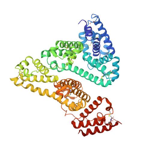
    3TDL

  • PubMed Abstract: 

    11-(Dansylamino) undecanoic acid (DAUDA) is a dansyl-type fluorophore and has widely used as a probe to determine the binding site for human serum albumin (HSA). Here, we reported that structure of HSA-Myristate-DAUDA ternary complex and identified clearly the presence of two DAUDA molecules at fatty acid (FA) binding site 6 and 7 of HSA, thus showing these two sites are weak FA binding sites. This result also show that DAUDA is an appropriate probe for FA site 6 and 7 on HSA as previous studied, but not a good probe of FA binding site 1 that is likely bilirubin binding site on HSA.


  • Organizational Affiliation

    State Key Laboratory of Structural Chemistry, Fujian Institute of Research on the Structure of Matter, Chinese Academy of Sciences, Fuzhou, China.


Macromolecules
Find similar proteins by:  (by identity cutoff)  |  3D Structure
Entity ID: 1
MoleculeChains Sequence LengthOrganismDetailsImage
Serum albumin585Homo sapiensMutation(s): 0 
Gene Names: ALBGIG20GIG42PRO0903PRO1708PRO2044PRO2619PRO2675UNQ696/PRO1341
UniProt & NIH Common Fund Data Resources
Find proteins for P02768 (Homo sapiens)
Explore P02768 
Go to UniProtKB:  P02768
PHAROS:  P02768
GTEx:  ENSG00000163631 
Entity Groups  
Sequence Clusters30% Identity50% Identity70% Identity90% Identity95% Identity100% Identity
UniProt GroupP02768
Sequence Annotations
Expand
  • Reference Sequence
Experimental Data & Validation

Experimental Data

  • Method: X-RAY DIFFRACTION
  • Resolution: 2.60 Å
  • R-Value Free:  0.261 (Depositor), 0.300 (DCC) 
  • R-Value Work:  0.251 (Depositor), 0.240 (DCC) 
  • R-Value Observed: 0.251 (Depositor) 
Space Group: C 1 2 1
Unit Cell:
Length ( Å )Angle ( ˚ )
a = 176.657α = 90
b = 37.137β = 104.38
c = 90.195γ = 90
Software Package:
Software NamePurpose
CNSrefinement
MAR345dtbdata collection
HKL-2000data reduction
HKL-2000data scaling
CNSphasing

Structure Validation

View Full Validation Report


Ligand Structure Quality Assessment 

Created with Raphaël 2.3.0Worse 01 BetterLigand structure goodness of fit to experimental dataBest fitted 11DClick on this verticalbar to view detailsBest fitted MYRClick on this verticalbar to view details

Entry History 

Deposition Data

Revision History  (Full details and data files)

  • Version 1.0: 2012-06-27
    Type: Initial release
  • Version 1.1: 2023-11-01
    Changes: Data collection, Database references, Derived calculations, Refinement description
  • Version 1.2: 2024-11-13
    Changes: Structure summary