3TN0

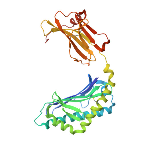
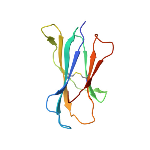
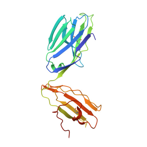
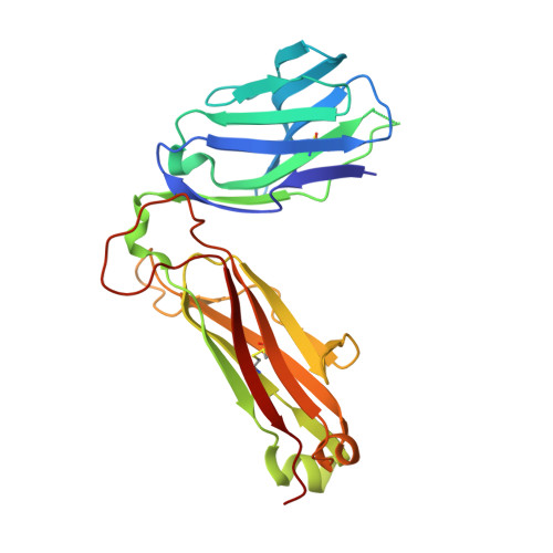
Structure of mouse Va14Vb8.2NKT TCR-mouse CD1d-a-C-Galactosylceramide complex


Experimental Data Snapshot

  • Method: X-RAY DIFFRACTION
  • Resolution: 3.20 Å
  • R-Value Free: 0.289 
  • R-Value Work: 0.225 
  • R-Value Observed: 0.229 

Starting Model: experimental
View more details

wwPDB Validation   3D Report Full Report


Ligand Structure Quality Assessment 


This is version 2.2 of the entry. See complete history


Literature

NKT TCR Recognition of CD1d-{alpha}-C-Galactosylceramide.

Patel, O.Cameron, G.Pellicci, D.G.Liu, Z.Byun, H.S.Beddoe, T.McCluskey, J.Franck, R.W.Castano, A.R.Harrak, Y.Llebaria, A.Bittman, R.Porcelli, S.A.Godfrey, D.I.Rossjohn, J.

(2011) J Immunol 187: 4705-4713

  • DOI: https://doi.org/10.4049/jimmunol.1100794
  • Primary Citation of Related Structures:  
    3TN0

  • PubMed Abstract: 

    NKT cells respond to a variety of CD1d-restricted glycolipid Ags that are structurally related to the prototypic Ag α-galactosylceramide (α-GalCer). A modified analog of α-GalCer with a carbon-based glycosidic linkage (α-C-GalCer) has generated great interest because of its apparent ability to promote prolonged, Th1-biased immune responses. In this study, we report the activation of spleen NKT cells to α-C-GalCer, and related C-glycoside ligands, is weaker than that of α-GalCer. Furthermore, the Vβ8.2 and Vβ7 NKT TCR affinity for CD1d-α-C-GalCer, and some related analogs, is ∼10-fold lower than that for the NKT TCR-CD1d-α-GalCer interaction. Nevertheless, the crystal structure of the Vβ8.2 NKT TCR-CD1d-α-C-GalCer complex is similar to that of the corresponding NKT TCR-CD1d-α-GalCer complex, although subtle differences at the interface provide a basis for understanding the lower affinity of the NKT TCR-CD1d-α-C-GalCer interaction. Our findings support the concept that for CD1d-restricted NKT cells, altered glycolipid ligands can promote markedly different responses while adopting similar TCR-docking topologies.


  • Organizational Affiliation

    Australian Research Council Centre of Excellence in Structural and Functional Microbial Genomics, Department of Biochemistry and Molecular Biology, School of Biomedical Sciences, Monash University, Clayton, Victoria 3800, Australia.


Macromolecules
Find similar proteins by:  (by identity cutoff)  |  3D Structure
Entity ID: 1
MoleculeChains Sequence LengthOrganismDetailsImage
Antigen-presenting glycoprotein CD1d1302Mus musculusMutation(s): 0 
Gene Names: Cd1d1Cd1.1
UniProt & NIH Common Fund Data Resources
Find proteins for P11609 (Mus musculus)
Go to UniProtKB:  P11609
IMPC:  MGI:107674
Glycosylation
Glycosylation Sites: 3Go to GlyGen: P11609-1
Sequence Annotations
Expand
  • Reference Sequence
Find similar proteins by:  (by identity cutoff)  |  3D Structure
Entity ID: 2
MoleculeChains Sequence LengthOrganismDetailsImage
Beta-2 microglobulin99Mus musculusMutation(s): 0 
Gene Names: B2mRP23-34E24.5-001mCG_11606
UniProt & NIH Common Fund Data Resources
Find proteins for P01887 (Mus musculus)
Go to UniProtKB:  P01887
IMPC:  MGI:88127
Sequence Annotations
Expand
  • Reference Sequence
Find similar proteins by:  (by identity cutoff)  |  3D Structure
Entity ID: 3
MoleculeChains Sequence LengthOrganismDetailsImage
mouse NKT Valpha14 (MOUSE VARIABLE DOMAIN, HUMAN CONSTANT DOMAIN)207Homo sapiensMus musculus
This entity is chimeric
Mutation(s): 0 
UniProt & NIH Common Fund Data Resources
Find proteins for P01848 (Homo sapiens)
Go to UniProtKB:  P01848
PHAROS:  P01848
Sequence Annotations
Expand
  • Reference Sequence
Find similar proteins by:  (by identity cutoff)  |  3D Structure
Entity ID: 4
MoleculeChains Sequence LengthOrganismDetailsImage
mouse NKT Vbeta8.2 (MOUSE VARIABLE DOMAIN, HUMAN CONSTANT DOMAIN)244Homo sapiensMus musculus
This entity is chimeric
Mutation(s): 0 
UniProt
Find proteins for P01850 (Homo sapiens)
Go to UniProtKB:  P01850
Sequence Annotations
Expand
  • Reference Sequence
Oligosaccharides

Help

Entity ID: 5
MoleculeChains Length2D Diagram Glycosylation3D Interactions
2-acetamido-2-deoxy-beta-D-glucopyranose-(1-4)-2-acetamido-2-deoxy-beta-D-glucopyranose
E
2N-Glycosylation
Glycosylation Resources
GlyTouCan:  G42666HT
GlyCosmos:  G42666HT
GlyGen:  G42666HT
Small Molecules
Ligands 2 Unique
IDChains Name / Formula / InChI Key2D Diagram3D Interactions
QUX
Query on QUX

Download Ideal Coordinates CCD File 
F [auth A]N-[(3S,4S,5R)-4,5-dihydroxy-1-[(2R,3R,4R,5R,6R)-3,4,5-trihydroxy-6-(hydroxymethyl)oxan-2-yl]nonadecan-3-yl]hexacosanamide
C51 H101 N O8
BNYLLEHBKIGJHB-XHSUSRKPSA-N
NAG
Query on NAG

Download Ideal Coordinates CCD File 
G [auth A],
H [auth A]
2-acetamido-2-deoxy-beta-D-glucopyranose
C8 H15 N O6
OVRNDRQMDRJTHS-FMDGEEDCSA-N
Binding Affinity Annotations 
IDSourceBinding Affinity
QUX PDBBind:  3TN0 Kd: 2200 (nM) from 1 assay(s)
Experimental Data & Validation

Experimental Data

  • Method: X-RAY DIFFRACTION
  • Resolution: 3.20 Å
  • R-Value Free: 0.289 
  • R-Value Work: 0.225 
  • R-Value Observed: 0.229 
  • Space Group: P 21 21 21
Unit Cell:
Length ( Å )Angle ( ˚ )
a = 59.171α = 90
b = 86.368β = 90
c = 236.403γ = 90
Software Package:
Software NamePurpose
Blu-Icedata collection
PHASERphasing
REFMACrefinement
MOSFLMdata reduction
SCALAdata scaling

Structure Validation

View Full Validation Report


Ligand Structure Quality Assessment 


Entry History 

Deposition Data

Revision History  (Full details and data files)

  • Version 1.0: 2011-12-14
    Type: Initial release
  • Version 2.0: 2020-07-29
    Type: Remediation
    Reason: Carbohydrate remediation
    Changes: Advisory, Atomic model, Data collection, Database references, Derived calculations, Structure summary
  • Version 2.1: 2023-09-13
    Changes: Advisory, Data collection, Database references, Refinement description, Structure summary
  • Version 2.2: 2024-10-30
    Changes: Structure summary