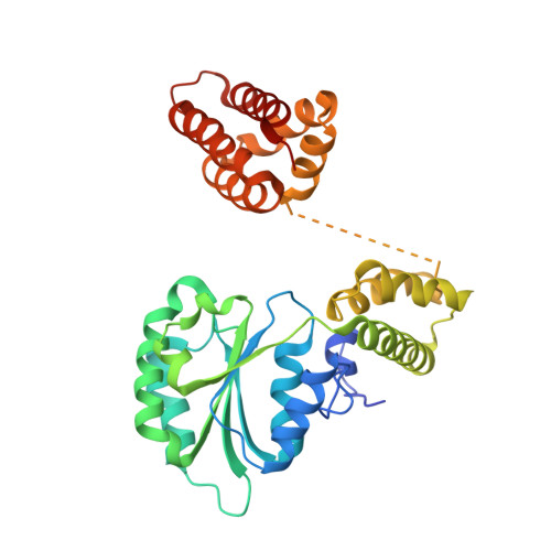
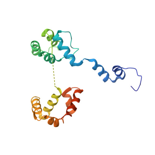
3U61

Structure of T4 Bacteriophage Clamp Loader Bound To Closed Clamp, DNA and ATP Analog and ADP


Experimental Data Snapshot

  • Method: X-RAY DIFFRACTION
  • Resolution: 3.20 Å
  • R-Value Free: 0.305 
  • R-Value Work: 0.239 
  • R-Value Observed: 0.244 

wwPDB Validation   3D Report Full Report


Ligand Structure Quality Assessment 


This is version 1.1 of the entry. See complete history


Literature

How a DNA polymerase clamp loader opens a sliding clamp.

Kelch, B.A.Makino, D.L.O'Donnell, M.Kuriyan, J.

(2011) Science 334: 1675-1680

  • DOI: https://doi.org/10.1126/science.1211884
  • Primary Citation of Related Structures:  
    3U5Z, 3U60, 3U61

  • PubMed Abstract: 

    Processive chromosomal replication relies on sliding DNA clamps, which are loaded onto DNA by pentameric clamp loader complexes belonging to the AAA+ family of adenosine triphosphatases (ATPases). We present structures for the ATP-bound state of the clamp loader complex from bacteriophage T4, bound to an open clamp and primer-template DNA. The clamp loader traps a spiral conformation of the open clamp so that both the loader and the clamp match the helical symmetry of DNA. One structure reveals that ATP has been hydrolyzed in one subunit and suggests that clamp closure and ejection of the loader involves disruption of the ATP-dependent match in symmetry. The structures explain how synergy among the loader, the clamp, and DNA can trigger ATP hydrolysis and release of the closed clamp on DNA.


  • Organizational Affiliation

    Department of Molecular and Cell Biology and California Institute for Quantitative Biosciences, University of California, Berkeley, CA 94720, USA.


Macromolecules

Find similar proteins by:  (by identity cutoff)  |  3D Structure
Entity ID: 1
MoleculeChains Sequence LengthOrganismDetailsImage
DNA polymerase accessory protein 44A [auth B],
B [auth C],
C [auth D],
D [auth E]
324Tequatrovirus T4Mutation(s): 0 
Gene Names: 44gp44
EC: 3.6.4
UniProt
Find proteins for P04526 (Enterobacteria phage T4)
Explore P04526 
Go to UniProtKB:  P04526
Entity Groups  
Sequence Clusters30% Identity50% Identity70% Identity90% Identity95% Identity100% Identity
UniProt GroupP04526
Sequence Annotations
Expand
  • Reference Sequence
Find similar proteins by:  (by identity cutoff)  |  3D Structure
Entity ID: 2
MoleculeChains Sequence LengthOrganismDetailsImage
DNA polymerase accessory protein 62E [auth A]199Tequatrovirus T4Mutation(s): 0 
Gene Names: 62gp62
UniProt
Find proteins for P04527 (Enterobacteria phage T4)
Explore P04527 
Go to UniProtKB:  P04527
Entity Groups  
Sequence Clusters30% Identity50% Identity70% Identity90% Identity95% Identity100% Identity
UniProt GroupP04527
Sequence Annotations
Expand
  • Reference Sequence
Find similar proteins by:  (by identity cutoff)  |  3D Structure
Entity ID: 3
MoleculeChains Sequence LengthOrganismDetailsImage
DNA polymerase processivity componentF [auth G],
G [auth H],
H [auth F]
228Tequatrovirus T4Mutation(s): 0 
Gene Names: 45gp45
UniProt
Find proteins for P04525 (Enterobacteria phage T4)
Explore P04525 
Go to UniProtKB:  P04525
Entity Groups  
Sequence Clusters30% Identity50% Identity70% Identity90% Identity95% Identity100% Identity
UniProt GroupP04525
Sequence Annotations
Expand
  • Reference Sequence

Find similar nucleic acids by:  Sequence   |   3D Structure  

Entity ID: 4
MoleculeChains LengthOrganismImage
Template DNA strand20N/A
Sequence Annotations
Expand
  • Reference Sequence

Find similar nucleic acids by:  Sequence   |   3D Structure  

Entity ID: 5
MoleculeChains LengthOrganismImage
Primer DNA strand10N/A
Sequence Annotations
Expand
  • Reference Sequence
Small Molecules
Ligands 3 Unique
IDChains Name / Formula / InChI Key2D Diagram3D Interactions
08T
Query on 08T

Download Ideal Coordinates CCD File 
M [auth C],
O [auth D]
[[[(2R,3S,4R,5R)-5-(6-aminopurin-9-yl)-3,4-bis(oxidanyl)oxolan-2-yl]methoxy-oxidanyl-phosphoryl]oxy-oxidanyl-phosphoryl]oxy-tris(fluoranyl)beryllium
C10 H14 Be F3 N5 O10 P2
WOGYHYSOODLXII-KWIZKVQNSA-J
ADP
Query on ADP

Download Ideal Coordinates CCD File 
K [auth B]ADENOSINE-5'-DIPHOSPHATE
C10 H15 N5 O10 P2
XTWYTFMLZFPYCI-KQYNXXCUSA-N
MG
Query on MG

Download Ideal Coordinates CCD File 
L [auth B],
N [auth C],
P [auth D]
MAGNESIUM ION
Mg
JLVVSXFLKOJNIY-UHFFFAOYSA-N
Modified Residues  1 Unique
IDChains TypeFormula2D DiagramParent
MSE
Query on MSE
F [auth G],
G [auth H],
H [auth F]
L-PEPTIDE LINKINGC5 H11 N O2 SeMET
Experimental Data & Validation

Experimental Data

  • Method: X-RAY DIFFRACTION
  • Resolution: 3.20 Å
  • R-Value Free: 0.305 
  • R-Value Work: 0.239 
  • R-Value Observed: 0.244 
  • Space Group: P 21 21 21
Unit Cell:
Length ( Å )Angle ( ˚ )
a = 82.303α = 90
b = 137.272β = 90
c = 222.779γ = 90
Software Package:
Software NamePurpose
HKL-2000data collection
PHASERphasing
PHENIXrefinement
HKL-2000data reduction
HKL-2000data scaling

Structure Validation

View Full Validation Report


Ligand Structure Quality Assessment 


Entry History 

Deposition Data

Revision History  (Full details and data files)

  • Version 1.0: 2012-01-04
    Type: Initial release
  • Version 1.1: 2024-10-30
    Changes: Data collection, Database references, Derived calculations, Structure summary