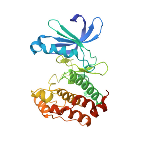
3UNZ

Aurora A in Complex with RPM1679


Experimental Data Snapshot

  • Method: X-RAY DIFFRACTION
  • Resolution: 2.80 Å
  • R-Value Free: 0.278 
  • R-Value Work: 0.231 

Starting Model: experimental
View more details

wwPDB Validation   3D Report Full Report


Ligand Structure Quality Assessment 


This is version 1.2 of the entry. See complete history


Literature

A Novel Mechanism by Which Small Molecule Inhibitors Induce the DFG Flip in Aurora A.

Martin, M.P.Zhu, J.Y.Lawrence, H.R.Pireddu, R.Luo, Y.Alam, R.Ozcan, S.Sebti, S.M.Lawrence, N.J.Schonbrunn, E.

(2012) ACS Chem Biol 7: 698-706

  • DOI: https://doi.org/10.1021/cb200508b
  • Primary Citation of Related Structures:  
    3UNJ, 3UNK, 3UNZ, 3UO4, 3UO5, 3UO6, 3UOD, 3UOH, 3UOJ, 3UOK, 3UOL, 3UP2

  • PubMed Abstract: 

    Most protein kinases share a DFG (Asp-Phe-Gly) motif in the ATP site that can assume two distinct conformations, the active DFG-in and the inactive DFG-out states. Small molecule inhibitors able to induce the DFG-out state have received considerable attention in kinase drug discovery. Using a typical DFG-in inhibitor scaffold of Aurora A, a kinase involved in the regulation of cell division, we found that halogen and nitrile substituents directed at the N-terminally flanking residue Ala273 induced global conformational changes in the enzyme, leading to DFG-out inhibitors that are among the most potent Aurora A inhibitors reported to date. The data suggest an unprecedented mechanism of action, in which induced-dipole forces along the Ala273 side chain alter the charge distribution of the DFG backbone, allowing the DFG to unwind. As the ADFG sequence and three-dimensional structure is highly conserved, DFG-out inhibitors of other kinases may be designed by specifically targeting the flanking alanine residue with electric dipoles.


  • Organizational Affiliation

    Drug Discovery Department, Moffitt Cancer Center and Research Institute, Tampa, Florida 33612, United States.


Macromolecules
Find similar proteins by:  (by identity cutoff)  |  3D Structure
Entity ID: 1
MoleculeChains Sequence LengthOrganismDetailsImage
Aurora kinase A
A, B
279Homo sapiensMutation(s): 1 
Gene Names: AURKAAIKAIRK1ARK1AURAAYK1BTAKIAK1STK15STK6
EC: 2.7.11.1
UniProt & NIH Common Fund Data Resources
Find proteins for O14965 (Homo sapiens)
Go to UniProtKB:  O14965
PHAROS:  O14965
GTEx:  ENSG00000087586 
Sequence Annotations
Expand
  • Reference Sequence
Small Molecules
Binding Affinity Annotations 
IDSourceBinding Affinity
0BZ BindingDB:  3UNZ Kd: 16 (nM) from 1 assay(s)
IC50: 3.7 (nM) from 1 assay(s)
-TΔS: -2.59e+1 (kJ/mol) from 1 assay(s)
ΔH: -7.10e+1 (kJ/mol) from 1 assay(s)
PDBBind:  3UNZ Kd: 16 (nM) from 1 assay(s)
Experimental Data & Validation

Experimental Data

  • Method: X-RAY DIFFRACTION
  • Resolution: 2.80 Å
  • R-Value Free: 0.278 
  • R-Value Work: 0.231 
  • Space Group: P 32
Unit Cell:
Length ( Å )Angle ( ˚ )
a = 85.78α = 90
b = 85.78β = 90
c = 76.56γ = 120
Software Package:
Software NamePurpose
PHENIXmodel building
PHENIXrefinement
XDSdata reduction
XDSdata scaling
PHENIXphasing

Structure Validation

View Full Validation Report


Ligand Structure Quality Assessment 


Entry History 

Deposition Data

Revision History  (Full details and data files)

  • Version 1.0: 2012-01-25
    Type: Initial release
  • Version 1.1: 2012-05-02
    Changes: Database references
  • Version 1.2: 2023-09-13
    Changes: Data collection, Database references, Derived calculations, Refinement description