3VZC | pdb_00003vzc

Crystal structure of Sphingosine Kinase 1 with inhibitor


Experimental Data Snapshot

  • Method: X-RAY DIFFRACTION
  • Resolution: 2.30 Å
  • R-Value Free: 
    0.285 (Depositor), 0.280 (DCC) 
  • R-Value Work: 
    0.229 (Depositor), 0.230 (DCC) 
  • R-Value Observed: 
    0.232 (Depositor) 

wwPDB Validation   3D Report Full Report


Ligand Structure Quality Assessment 

Created with Raphaël 2.3.0Worse 01 BetterLigand structure goodness of fit to experimental dataBest fitted UULClick on this verticalbar to view details

This is version 1.2 of the entry. See complete history


Literature

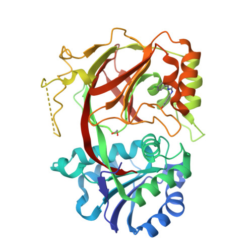
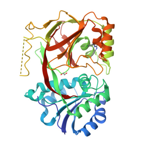
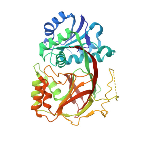
Molecular basis of sphingosine kinase 1 substrate recognition and catalysis.

Wang, Z.Min, X.Xiao, S.H.Johnstone, S.Romanow, W.Meininger, D.Xu, H.Liu, J.Dai, J.An, S.Thibault, S.Walker, N.

(2013) Structure 21: 798-809

  • DOI: https://doi.org/10.1016/j.str.2013.02.025
  • Primary Citation of Related Structures:  
    3VZB, 3VZC, 3VZD

  • PubMed Abstract: 

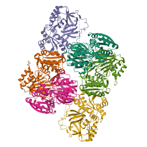
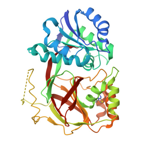
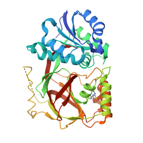
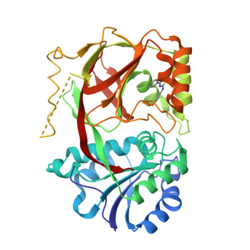
    Sphingosine kinase 1 (SphK1) is a lipid kinase that catalyzes the conversion of sphingosine to sphingosine-1-phosphate (S1P), which has been shown to play a role in lymphocyte trafficking, angiogenesis, and response to apoptotic stimuli. As a central enzyme in modulating the S1P levels in cells, SphK1 emerges as an important regulator for diverse cellular functions and a potential target for drug discovery. Here, we present the crystal structures of human SphK1 in the apo form and in complexes with a substrate sphingosine-like lipid, ADP, and an inhibitor at 2.0-2.3 Å resolution. The SphK1 structures reveal a two-domain architecture in which its catalytic site is located in the cleft between the two domains and a hydrophobic lipid-binding pocket is buried in the C-terminal domain. Comparative analysis of these structures with mutagenesis and kinetic studies provides insight into how SphK1 recognizes the lipid substrate and catalyzes ATP-dependent phosphorylation.


  • Organizational Affiliation

    Department of Molecular Structure and Characterization, Amgen, Inc., 1120 Veterans Boulevard, South San Francisco, CA 94080, USA. zwang@amgen.com


Macromolecules
Find similar proteins by:  (by identity cutoff)  |  3D Structure
Entity ID: 1
MoleculeChains Sequence LengthOrganismDetailsImage
Sphingosine kinase 1
A, B, C, D, E
361Homo sapiensMutation(s): 0 
Gene Names: SPHK1SPHKSPK
EC: 2.7.1.91 (PDB Primary Data), 2.3.1 (UniProt)
UniProt & NIH Common Fund Data Resources
Find proteins for Q9NYA1 (Homo sapiens)
Explore Q9NYA1 
Go to UniProtKB:  Q9NYA1
PHAROS:  Q9NYA1
GTEx:  ENSG00000176170 
Entity Groups  
Sequence Clusters30% Identity50% Identity70% Identity90% Identity95% Identity100% Identity
UniProt GroupQ9NYA1
Sequence Annotations
Expand
  • Reference Sequence
Small Molecules
Ligands 2 Unique
IDChains Name / Formula / InChI Key2D Diagram3D Interactions
UUL
Query on UUL

Download Ideal Coordinates CCD File 
G [auth A]
H [auth B]
I [auth C]
J [auth D]
L [auth E]
4-{[4-(4-chlorophenyl)-1,3-thiazol-2-yl]amino}phenol
C15 H11 Cl N2 O S
ZFGXZJKLOFCECI-UHFFFAOYSA-N
EDO
Query on EDO

Download Ideal Coordinates CCD File 
K [auth D],
N [auth F]
1,2-ETHANEDIOL
C2 H6 O2
LYCAIKOWRPUZTN-UHFFFAOYSA-N
Binding Affinity Annotations 
IDSourceBinding Affinity
UUL BindingDB:  3VZC Ki: min: 500, max: 1.70e+4 (nM) from 4 assay(s)
IC50: min: 500, max: 3.50e+4 (nM) from 2 assay(s)
Experimental Data & Validation

Experimental Data

  • Method: X-RAY DIFFRACTION
  • Resolution: 2.30 Å
  • R-Value Free:  0.285 (Depositor), 0.280 (DCC) 
  • R-Value Work:  0.229 (Depositor), 0.230 (DCC) 
  • R-Value Observed: 0.232 (Depositor) 
Space Group: P 21 21 21
Unit Cell:
Length ( Å )Angle ( ˚ )
a = 102.22α = 90
b = 106.61β = 90
c = 226.18γ = 90
Software Package:
Software NamePurpose
SCALAdata scaling
REFMACrefinement
PDB_EXTRACTdata extraction

Structure Validation

View Full Validation Report


Ligand Structure Quality Assessment 

Created with Raphaël 2.3.0Worse 01 BetterLigand structure goodness of fit to experimental dataBest fitted UULClick on this verticalbar to view details

Entry History 

Deposition Data

Revision History  (Full details and data files)

  • Version 1.0: 2013-05-08
    Type: Initial release
  • Version 1.1: 2014-01-29
    Changes: Database references
  • Version 1.2: 2024-03-20
    Changes: Data collection, Database references, Derived calculations