3AB4

Crystal structure of feedback inhibition resistant mutant of aspartate kinase from Corynebacterium glutamicum in complex with lysine and threonine


Experimental Data Snapshot

  • Method: X-RAY DIFFRACTION
  • Resolution: 2.47 Å
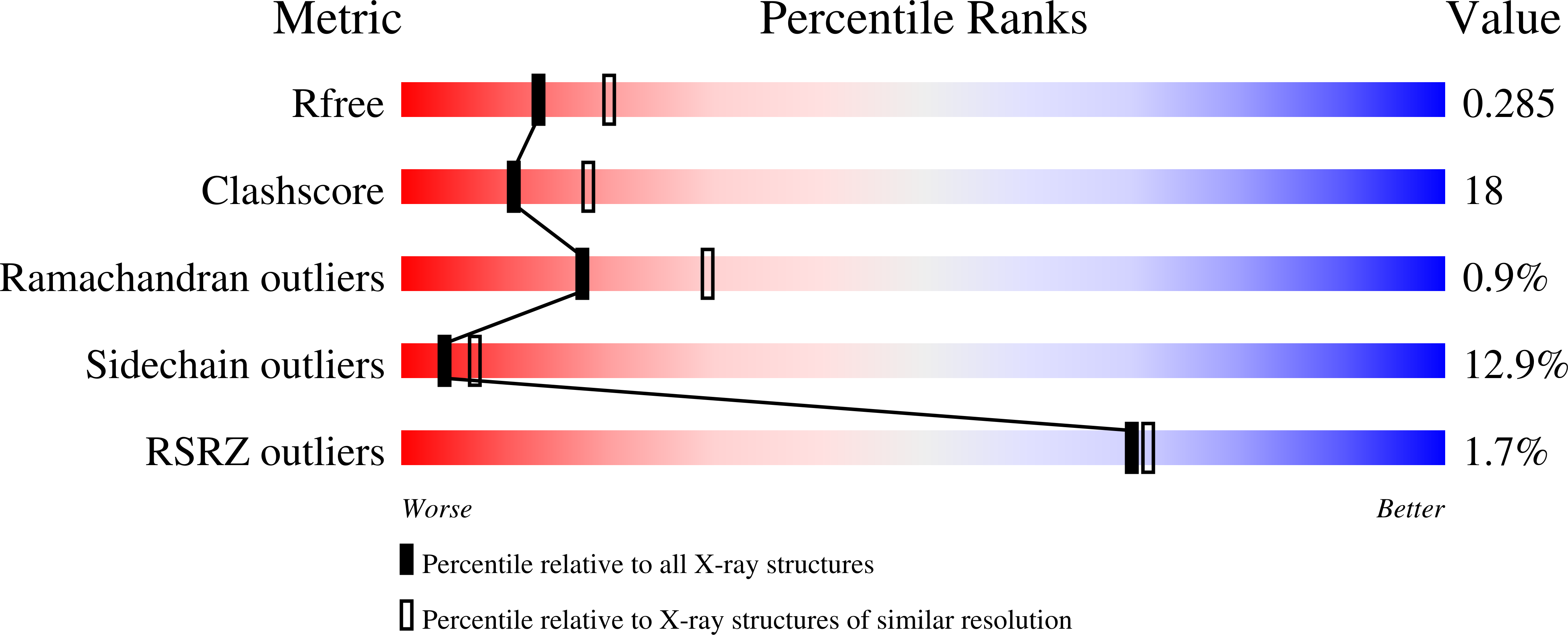
  • R-Value Free: 0.288 
  • R-Value Work: 0.223 
  • R-Value Observed: 0.226 

Starting Model: experimental
View more details

wwPDB Validation   3D Report Full Report


This is version 1.2 of the entry. See complete history


Literature

Mechanism of concerted inhibition of {alpha}2{beta}2-type heterooligomeric aspartate kinase from Corynebacterium glutamicum

Yoshida, A.Tomita, T.Kuzuyama, T.Nishiyama, M.

(2010) J Biol Chem 285: 27477-27486

  • DOI: https://doi.org/10.1074/jbc.M110.111153
  • Primary Citation of Related Structures:  
    3AAW, 3AB2, 3AB4

  • PubMed Abstract: 

    Aspartate kinase (AK) is the first and committed enzyme of the biosynthetic pathway producing aspartate family amino acids, lysine, threonine, and methionine. AK from Corynebacterium glutamicum (CgAK), a bacterium used for industrial fermentation of amino acids, including glutamate and lysine, is inhibited by lysine and threonine in a concerted manner. To elucidate the mechanism of this unique regulation in CgAK, we determined the crystal structures in several forms: an inhibitory form complexed with both lysine and threonine, an active form complexed with only threonine, and a feedback inhibition-resistant mutant (S301F) complexed with both lysine and threonine. CgAK has a characteristic alpha(2)beta(2)-type heterotetrameric structure made up of two alpha subunits and two beta subunits. Comparison of the crystal structures between inhibitory and active forms revealed that binding inhibitors causes a conformational change to a closed inhibitory form, and the interaction between the catalytic domain in the alpha subunit and beta subunit (regulatory subunit) is a key event for stabilizing the inhibitory form. This study shows not only the first crystal structures of alpha(2)beta(2)-type AK but also the mechanism of concerted inhibition in CgAK.


  • Organizational Affiliation

    Biotechnology Research Center, The University of Tokyo, 1-1-1 Yayoi, Bunkyo-ku, Tokyo 113-8657, Japan.


Macromolecules
Find similar proteins by:  (by identity cutoff)  |  3D Structure
Entity ID: 1
MoleculeChains Sequence LengthOrganismDetailsImage
Aspartokinase
A, C, E, G, I
A, C, E, G, I, K, M, O
421Corynebacterium glutamicumMutation(s): 0 
Gene Names: lysC
EC: 2.7.2.4
UniProt
Find proteins for P26512 (Corynebacterium glutamicum (strain ATCC 13032 / DSM 20300 / JCM 1318 / BCRC 11384 / CCUG 27702 / LMG 3730 / NBRC 12168 / NCIMB 10025 / NRRL B-2784 / 534))
Go to UniProtKB:  P26512
Sequence Annotations
Expand
  • Reference Sequence
Find similar proteins by:  (by identity cutoff)  |  3D Structure
Entity ID: 2
MoleculeChains Sequence LengthOrganismDetailsImage
Aspartokinase
B, D, F, H, J
B, D, F, H, J, L, N, P
178Corynebacterium glutamicumMutation(s): 0 
Gene Names: lysC
EC: 2.7.2.4
UniProt
Find proteins for P26512 (Corynebacterium glutamicum (strain ATCC 13032 / DSM 20300 / JCM 1318 / BCRC 11384 / CCUG 27702 / LMG 3730 / NBRC 12168 / NCIMB 10025 / NRRL B-2784 / 534))
Go to UniProtKB:  P26512
Sequence Annotations
Expand
  • Reference Sequence
Small Molecules
Ligands 2 Unique
IDChains Name / Formula / InChI Key2D Diagram3D Interactions
LYS
Query on LYS

Download Ideal Coordinates CCD File 
AA [auth G]
FA [auth K]
IA [auth M]
LA [auth O]
R [auth A]
AA [auth G],
FA [auth K],
IA [auth M],
LA [auth O],
R [auth A],
U [auth C],
X [auth E]
LYSINE
C6 H15 N2 O2
KDXKERNSBIXSRK-YFKPBYRVSA-O
THR
Query on THR

Download Ideal Coordinates CCD File 
BA [auth H]
CA [auth I]
DA [auth J]
EA [auth K]
GA [auth L]
BA [auth H],
CA [auth I],
DA [auth J],
EA [auth K],
GA [auth L],
HA [auth M],
JA [auth N],
KA [auth O],
MA [auth P],
Q [auth A],
S [auth B],
T [auth C],
V [auth D],
W [auth E],
Y [auth F],
Z [auth G]
THREONINE
C4 H9 N O3
AYFVYJQAPQTCCC-GBXIJSLDSA-N
Experimental Data & Validation

Experimental Data

  • Method: X-RAY DIFFRACTION
  • Resolution: 2.47 Å
  • R-Value Free: 0.288 
  • R-Value Work: 0.223 
  • R-Value Observed: 0.226 
  • Space Group: P 1
Unit Cell:
Length ( Å )Angle ( ˚ )
a = 99.033α = 76.03
b = 112.874β = 71.07
c = 120.009γ = 74.5
Software Package:
Software NamePurpose
HKL-2000data collection
MOLREPphasing
REFMACrefinement
HKL-2000data reduction
HKL-2000data scaling

Structure Validation

View Full Validation Report


Entry History 

Deposition Data

Revision History  (Full details and data files)

  • Version 1.0: 2010-06-23
    Type: Initial release
  • Version 1.1: 2011-07-13
    Changes: Version format compliance
  • Version 1.2: 2023-11-01
    Changes: Data collection, Database references, Derived calculations, Refinement description