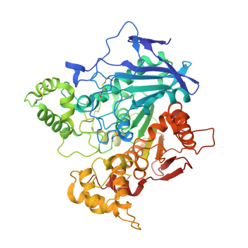
3DKK

Aged Form of Human Butyrylcholinesterase Inhibited by Tabun


Experimental Data Snapshot

  • Method: X-RAY DIFFRACTION
  • Resolution: 2.31 Å
  • R-Value Free: 0.251 
  • R-Value Work: 0.197 
  • R-Value Observed: 0.200 

wwPDB Validation   3D Report Full Report


Ligand Structure Quality Assessment 


This is version 2.1 of the entry. See complete history


Literature

Aging of Cholinesterases Phosphylated by Tabun Proceeds through O-Dealkylation.

Carletti, E.Li, H.Li, B.Ekstrom, F.Nicolet, Y.Loiodice, M.Gillon, E.Froment, M.T.Lockridge, O.Schopfer, L.M.Masson, P.Nachon, F.

(2008) J Am Chem Soc 130: 16011-16020

  • DOI: https://doi.org/10.1021/ja804941z
  • Primary Citation of Related Structures:  
    3DJY, 3DKK, 3DL4, 3DL7

  • PubMed Abstract: 

    Human butyrylcholinesterase (hBChE) hydrolyzes or scavenges a wide range of toxic esters, including heroin, cocaine, carbamate pesticides, organophosphorus pesticides, and nerve agents. Organophosphates (OPs) exert their acute toxicity through inhibition of acetylcholinesterase (AChE) by phosphorylation of the catalytic serine. Phosphylated cholinesterase (ChE) can undergo a spontaneous, time-dependent process called "aging", during which the OP-ChE conjugate is dealkylated. This leads to irreversible inhibition of the enzyme. The inhibition of ChEs by tabun and the subsequent aging reaction are of particular interest, because tabun-ChE conjugates display an extraordinary resistance toward most current oxime reactivators. We investigated the structural basis of oxime resistance for phosphoramidated ChE conjugates by determining the crystal structures of the non-aged and aged forms of hBChE inhibited by tabun, and by updating the refinement of non-aged and aged tabun-inhibited mouse AChE (mAChE). Structures for non-aged and aged tabun-hBChE were refined to 2.3 and 2.1 A, respectively. The refined structures of aged ChE conjugates clearly show that the aging reaction proceeds through O-dealkylation of the P(R) enantiomer of tabun. After dealkylation, the negatively charged oxygen forms a strong salt bridge with protonated His438N epsilon2 that prevents reactivation. Mass spectrometric analysis of the aged tabun-inhibited hBChE showed that both the dimethylamine and ethoxy side chains were missing from the phosphorus. Loss of the ethoxy is consistent with the crystallography results. Loss of the dimethylamine is consistent with acid-catalyzed deamidation during the preparation of the aged adduct for mass spectrometry. The reported 3D data will help in the design of new oximes capable of reactivating tabun-ChE conjugates.


  • Organizational Affiliation

    Département de Toxicologie, Centre de Recherches du Service de Santé des Armées, 38700 La Tronche, France.


Macromolecules
Find similar proteins by:  (by identity cutoff)  |  3D Structure
Entity ID: 1
MoleculeChains Sequence LengthOrganismDetailsImage
Cholinesterase529Homo sapiensMutation(s): 4 
Gene Names: BCHECHE1
EC: 3.1.1.8
UniProt & NIH Common Fund Data Resources
Find proteins for P06276 (Homo sapiens)
Explore P06276 
Go to UniProtKB:  P06276
PHAROS:  P06276
GTEx:  ENSG00000114200 
Entity Groups  
Sequence Clusters30% Identity50% Identity70% Identity90% Identity95% Identity100% Identity
UniProt GroupP06276
Glycosylation
Glycosylation Sites: 6Go to GlyGen: P06276-1
Sequence Annotations
Expand
  • Reference Sequence
Oligosaccharides

Help

Entity ID: 2
MoleculeChains Length2D Diagram Glycosylation3D Interactions
2-acetamido-2-deoxy-beta-D-glucopyranose-(1-4)-[beta-L-fucopyranose-(1-6)]2-acetamido-2-deoxy-beta-D-glucopyranose
B, D
3N-Glycosylation
Glycosylation Resources
GlyTouCan:  G28454KX
GlyCosmos:  G28454KX
GlyGen:  G28454KX
Entity ID: 3
MoleculeChains Length2D Diagram Glycosylation3D Interactions
beta-L-fucopyranose-(1-6)-2-acetamido-2-deoxy-beta-D-glucopyranose
C
2N-Glycosylation
Glycosylation Resources
GlyTouCan:  G61843VN
GlyCosmos:  G61843VN
GlyGen:  G61843VN
Small Molecules
Ligands 4 Unique
IDChains Name / Formula / InChI Key2D Diagram3D Interactions
NAG
Query on NAG

Download Ideal Coordinates CCD File 
E [auth A],
F [auth A],
G [auth A]
2-acetamido-2-deoxy-beta-D-glucopyranose
C8 H15 N O6
OVRNDRQMDRJTHS-FMDGEEDCSA-N
SO4
Query on SO4

Download Ideal Coordinates CCD File 
K [auth A]SULFATE ION
O4 S
QAOWNCQODCNURD-UHFFFAOYSA-L
CL
Query on CL

Download Ideal Coordinates CCD File 
H [auth A],
I [auth A]
CHLORIDE ION
Cl
VEXZGXHMUGYJMC-UHFFFAOYSA-M
NA
Query on NA

Download Ideal Coordinates CCD File 
J [auth A]SODIUM ION
Na
FKNQFGJONOIPTF-UHFFFAOYSA-N
Modified Residues  1 Unique
IDChains TypeFormula2D DiagramParent
SEN
Query on SEN
A
L-PEPTIDE LINKINGC5 H13 N2 O5 PSER
Experimental Data & Validation

Experimental Data

  • Method: X-RAY DIFFRACTION
  • Resolution: 2.31 Å
  • R-Value Free: 0.251 
  • R-Value Work: 0.197 
  • R-Value Observed: 0.200 
  • Space Group: I 4 2 2
Unit Cell:
Length ( Å )Angle ( ˚ )
a = 155.24α = 90
b = 155.24β = 90
c = 127.47γ = 90
Software Package:
Software NamePurpose
XSCALEdata scaling
MOLREPphasing
REFMACrefinement
PDB_EXTRACTdata extraction
PROCDdata collection
XDSdata reduction

Structure Validation

View Full Validation Report


Ligand Structure Quality Assessment 


Entry History 

Deposition Data

Revision History  (Full details and data files)

  • Version 1.0: 2008-12-02
    Type: Initial release
  • Version 1.1: 2011-07-13
    Changes: Non-polymer description, Version format compliance
  • Version 2.0: 2020-07-29
    Type: Remediation
    Reason: Carbohydrate remediation
    Changes: Atomic model, Data collection, Derived calculations, Structure summary
  • Version 2.1: 2021-10-20
    Changes: Database references, Structure summary