3W37

Sugar beet alpha-glucosidase with acarbose


Experimental Data Snapshot

  • Method: X-RAY DIFFRACTION
  • Resolution: 1.70 Å
  • R-Value Free: 
    0.182 (Depositor), 0.179 (DCC) 
  • R-Value Work: 
    0.154 (Depositor), 0.151 (DCC) 
  • R-Value Observed: 
    0.154 (Depositor) 

wwPDB Validation   3D Report Full Report


Ligand Structure Quality Assessment 

Created with Raphaël 2.3.0Worse 01 BetterLigand structure goodness of fit to experimental dataBest fitted NAGClick on this verticalbar to view details

This is version 2.1 of the entry. See complete history


Literature

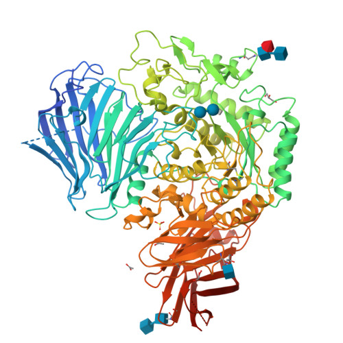
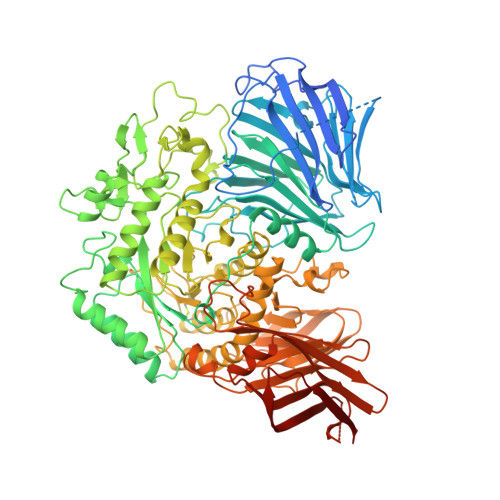
Molecular basis for the recognition of long-chain substrates by plant & alpha-glucosidase

Tagami, T.Yamashita, K.Okuyama, M.Mori, H.Yao, M.Kimura, A.

(2013) J Biol Chem 288: 19296-19303

  • DOI: https://doi.org/10.1074/jbc.M113.465211
  • Primary Citation of Related Structures:  
    3W37, 3W38

  • PubMed Abstract: 

    Sugar beet α-glucosidase (SBG), a member of glycoside hydrolase family 31, shows exceptional long-chain specificity, exhibiting higher kcat/Km values for longer malto-oligosaccharides. However, its amino acid sequence is similar to those of other short chain-specific α-glucosidases. To gain structural insights into the long-chain substrate recognition of SBG, a crystal structure complex with the pseudotetrasaccharide acarbose was determined at 1.7 Å resolution. The active site pocket of SBG is formed by a (β/α)8 barrel domain and a long loop (N-loop) bulging from the N-terminal domain similar to other related enzymes. Two residues (Phe-236 and Asn-237) in the N-loop are important for the long-chain specificity. Kinetic analysis of an Asn-237 mutant enzyme and a previous study of a Phe-236 mutant enzyme demonstrated that these residues create subsites +2 and +3. The structure also indicates that Phe-236 and Asn-237 guide the reducing end of long substrates to subdomain b2, which is an additional element inserted into the (β/α)8 barrel domain. Subdomain b2 of SBG includes Ser-497, which was identified as the residue at subsite +4 by site-directed mutagenesis.


  • Organizational Affiliation

    Research Faculty of Agriculture, Hokkaido University, Sapporo 060-8589, Japan.


Macromolecules
Find similar proteins by:  (by identity cutoff)  |  3D Structure
Entity ID: 1
MoleculeChains Sequence LengthOrganismDetailsImage
Alpha-glucosidase913Beta vulgarisMutation(s): 0 
EC: 3.2.1.20
UniProt
Find proteins for L0N7E5 (Beta vulgaris)
Explore L0N7E5 
Go to UniProtKB:  L0N7E5
Entity Groups  
Sequence Clusters30% Identity50% Identity70% Identity90% Identity95% Identity100% Identity
UniProt GroupL0N7E5
Glycosylation
Glycosylation Sites: 3
Sequence Annotations
Expand
  • Reference Sequence
Oligosaccharides

Help

Entity ID: 2
MoleculeChains Length2D Diagram Glycosylation3D Interactions
2-acetamido-2-deoxy-beta-D-glucopyranose-(1-4)-2-acetamido-2-deoxy-beta-D-glucopyranose
B
2N-Glycosylation
Glycosylation Resources
GlyTouCan:  G42666HT
GlyCosmos:  G42666HT
GlyGen:  G42666HT
Entity ID: 3
MoleculeChains Length2D Diagram Glycosylation3D Interactions
alpha-L-fucopyranose-(1-3)-[2-acetamido-2-deoxy-beta-D-glucopyranose-(1-4)]2-acetamido-2-deoxy-beta-D-glucopyranose
C
3N-Glycosylation
Glycosylation Resources
GlyTouCan:  G11971MR
GlyCosmos:  G11971MR
GlyGen:  G11971MR
Entity ID: 4
MoleculeChains Length2D Diagram Glycosylation3D Interactions
4,6-dideoxy-4-{[(1S,4R,5S,6S)-4,5,6-trihydroxy-3-(hydroxymethyl)cyclohex-2-en-1-yl]amino}-alpha-D-glucopyranose-(1-4)-alpha-D-glucopyranose-(1-4)-alpha-D-glucopyranose
D
3N/AN/A
Glycosylation Resources
GlyTouCan:  G66431MI
GlyCosmos:  G66431MI
Small Molecules
Ligands 4 Unique
IDChains Name / Formula / InChI Key2D Diagram3D Interactions
NAG
Query on NAG

Download Ideal Coordinates CCD File 
J [auth A]2-acetamido-2-deoxy-beta-D-glucopyranose
C8 H15 N O6
OVRNDRQMDRJTHS-FMDGEEDCSA-N
SO4
Query on SO4

Download Ideal Coordinates CCD File 
H [auth A]SULFATE ION
O4 S
QAOWNCQODCNURD-UHFFFAOYSA-L
GOL
Query on GOL

Download Ideal Coordinates CCD File 
F [auth A],
I [auth A],
L [auth A]
GLYCEROL
C3 H8 O3
PEDCQBHIVMGVHV-UHFFFAOYSA-N
ACY
Query on ACY

Download Ideal Coordinates CCD File 
E [auth A],
G [auth A],
K [auth A]
ACETIC ACID
C2 H4 O2
QTBSBXVTEAMEQO-UHFFFAOYSA-N
Biologically Interesting Molecules (External Reference) 1 Unique
Experimental Data & Validation

Experimental Data

  • Method: X-RAY DIFFRACTION
  • Resolution: 1.70 Å
  • R-Value Free:  0.182 (Depositor), 0.179 (DCC) 
  • R-Value Work:  0.154 (Depositor), 0.151 (DCC) 
  • R-Value Observed: 0.154 (Depositor) 
Space Group: P 21 21 21
Unit Cell:
Length ( Å )Angle ( ˚ )
a = 86.36α = 90
b = 98.2β = 90
c = 108.75γ = 90
Software Package:
Software NamePurpose
XSCALEdata scaling
PHASERphasing
PHENIXrefinement
PDB_EXTRACTdata extraction
BSSdata collection
XDSdata reduction

Structure Validation

View Full Validation Report


Ligand Structure Quality Assessment 

Created with Raphaël 2.3.0Worse 01 BetterLigand structure goodness of fit to experimental dataBest fitted NAGClick on this verticalbar to view details

Entry History 

Deposition Data

Revision History  (Full details and data files)

  • Version 1.0: 2013-05-29
    Type: Initial release
  • Version 1.1: 2013-06-12
    Changes: Database references
  • Version 1.2: 2013-07-17
    Changes: Database references
  • Version 1.3: 2014-07-30
    Changes: Other
  • Version 2.0: 2020-07-29
    Type: Remediation
    Reason: Carbohydrate remediation
    Changes: Atomic model, Data collection, Derived calculations, Non-polymer description, Structure summary
  • Version 2.1: 2024-10-30
    Changes: Data collection, Database references, Structure summary