Structural and Mutational Studies on Substrate Specificity and Catalysis of Salmonella typhimurium D-Cysteine Desulfhydrase.
Bharath, S.R., Bisht, S., Harijan, R.K., Savithri, H.S., Murthy, M.R.N.(2012) PLoS One 7: e36267-e36267
- PubMed: 22574144
- DOI: https://doi.org/10.1371/journal.pone.0036267
- Primary Citation of Related Structures:
4D8T, 4D8U, 4D8W, 4D92, 4D96, 4D97, 4D99, 4D9B, 4D9C, 4D9E, 4D9F - PubMed Abstract:
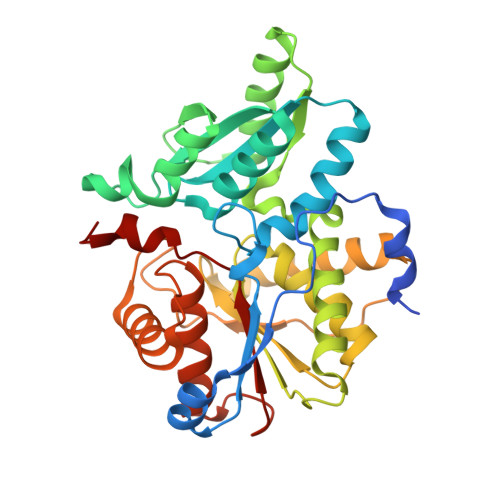
Salmonella typhimurium DCyD (StDCyD) is a fold type II pyridoxal 5' phosphate (PLP)-dependent enzyme that catalyzes the degradation of D-Cys to H(2)S and pyruvate. It also efficiently degrades β-chloro-D-alanine (βCDA). D-Ser is a poor substrate while the enzyme is inactive with respect to L-Ser and 1-amino-1-carboxy cyclopropane (ACC). Here, we report the X-ray crystal structures of StDCyD and of crystals obtained in the presence of D-Cys, βCDA, ACC, D-Ser, L-Ser, D-cycloserine (DCS) and L-cycloserine (LCS) at resolutions ranging from 1.7 to 2.6 Å. The polypeptide fold of StDCyD consisting of a small domain (residues 48-161) and a large domain (residues 1-47 and 162-328) resembles other fold type II PLP dependent enzymes. The structures obtained in the presence of D-Cys and βCDA show the product, pyruvate, bound at a site 4.0-6.0 Å away from the active site. ACC forms an external aldimine complex while D- and L-Ser bind non-covalently suggesting that the reaction with these ligands is arrested at Cα proton abstraction and transimination steps, respectively. In the active site of StDCyD cocrystallized with DCS or LCS, electron density for a pyridoxamine phosphate (PMP) was observed. Crystals soaked in cocktail containing these ligands show density for PLP-cycloserine. Spectroscopic observations also suggest formation of PMP by the hydrolysis of cycloserines. Mutational studies suggest that Ser78 and Gln77 are key determinants of enzyme specificity and the phenolate of Tyr287 is responsible for Cα proton abstraction from D-Cys. Based on these studies, a probable mechanism for the degradation of D-Cys by StDCyD is proposed.
Organizational Affiliation:
Molecular Biophysics Unit, Indian Institute of Science, Bangalore, India.


