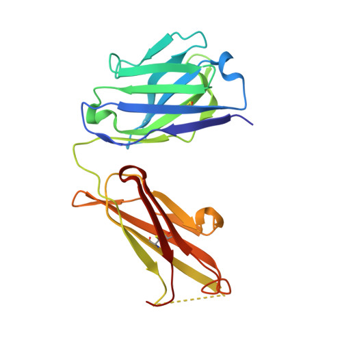
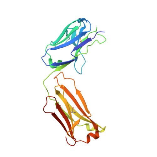
4DVB

The crystal structure of the Fab fragment of pro-uPA antibody mAb-112


Experimental Data Snapshot

  • Method: X-RAY DIFFRACTION
  • Resolution: 1.93 Å
  • R-Value Free: 0.259 
  • R-Value Work: 0.223 
  • R-Value Observed: 0.224 

wwPDB Validation   3D Report Full Report


This is version 1.0 of the entry. See complete history


Literature

Rezymogenation of active urokinase induced by an inhibitory antibody.

Jiang, L.Botkjaer, K.A.Andersen, L.M.Yuan, C.Andreasen, P.A.Huang, M.

(2013) Biochem J 449: 161-166

  • DOI: https://doi.org/10.1042/BJ20121132
  • Primary Citation of Related Structures:  
    4DVA, 4DVB, 4DW2

  • PubMed Abstract: 

    An important regulatory mechanism of serine proteases is the proteolytic conversion of the inactive pro-enzyme, or zymogen, into the active enzyme. This activation process is generally considered an irreversible process. In the present study, we demonstrate that an active enzyme can be converted back into its zymogen form. We determined the crystal structure of uPA (urokinase-type plasminogen activator) in complex with an inhibitory antibody, revealing that the antibody 'rezymogenizes' already activated uPA. The present study demonstrates a new regulatory mechanism of protease activity, which is also an extreme case of protein allostery. Mechanistically, the antibody binds a single surface-exposed loop, named the autolysis loop, thereby preventing the stabilization of uPA in its active conformation. We argue that this autolysis loop is a key structural element for rezymogenation of other proteases, and will be a new target site for pharmacological intervention with serine protease activity.


  • Organizational Affiliation

    State Key Laboratory of Structural Chemistry, Fujian Institute of Research on the Structure of Matter, Chinese Academy of Sciences, 155 Yang Qiao West Road, Fuzhou, Fujian, 350002, China.


Macromolecules
Find similar proteins by:  (by identity cutoff)  |  3D Structure
Entity ID: 1
MoleculeChains Sequence LengthOrganismDetailsImage
Fab fragment of pro-uPA antibody mAb-112A,
C [auth H]
213Mus musculusMutation(s): 0 
Entity Groups  
Sequence Clusters30% Identity50% Identity70% Identity90% Identity95% Identity100% Identity
Sequence Annotations
Expand
  • Reference Sequence
Find similar proteins by:  (by identity cutoff)  |  3D Structure
Entity ID: 2
MoleculeChains Sequence LengthOrganismDetailsImage
Fab fragment of pro-uPA antibody mAb-112B,
D [auth L]
215Mus musculusMutation(s): 0 
Entity Groups  
Sequence Clusters30% Identity50% Identity70% Identity90% Identity95% Identity100% Identity
Sequence Annotations
Expand
  • Reference Sequence
Small Molecules
Ligands 2 Unique
IDChains Name / Formula / InChI Key2D Diagram3D Interactions
PG4
Query on PG4

Download Ideal Coordinates CCD File 
H
TETRAETHYLENE GLYCOL
C8 H18 O5
UWHCKJMYHZGTIT-UHFFFAOYSA-N
SO4
Query on SO4

Download Ideal Coordinates CCD File 
E [auth A]
F [auth A]
G [auth B]
I [auth H]
J [auth H]
E [auth A],
F [auth A],
G [auth B],
I [auth H],
J [auth H],
K [auth H],
L
SULFATE ION
O4 S
QAOWNCQODCNURD-UHFFFAOYSA-L
Experimental Data & Validation

Experimental Data

  • Method: X-RAY DIFFRACTION
  • Resolution: 1.93 Å
  • R-Value Free: 0.259 
  • R-Value Work: 0.223 
  • R-Value Observed: 0.224 
  • Space Group: P 21 21 21
Unit Cell:
Length ( Å )Angle ( ˚ )
a = 71.177α = 90
b = 86.628β = 90
c = 155.331γ = 90
Software Package:
Software NamePurpose
ADSCdata collection
AMoREphasing
REFMACrefinement
HKL-2000data reduction
HKL-2000data scaling

Structure Validation

View Full Validation Report


Entry History 

Deposition Data

Revision History  (Full details and data files)

  • Version 1.0: 2013-01-16
    Type: Initial release