4EPP

Canonical poly(ADP-ribose) glycohydrolase from Tetrahymena thermophila.


Experimental Data Snapshot

  • Method: X-RAY DIFFRACTION
  • Resolution: 1.95 Å
  • R-Value Free: 
    0.207 (Depositor), 0.200 (DCC) 
  • R-Value Work: 
    0.167 (Depositor), 0.160 (DCC) 
  • R-Value Observed: 
    0.169 (Depositor) 

wwPDB Validation   3D Report Full Report


Ligand Structure Quality Assessment 

Created with Raphaël 2.3.0Worse 01 BetterLigand structure goodness of fit to experimental dataBest fitted APRClick on this verticalbar to view details

This is version 1.1 of the entry. See complete history


Literature

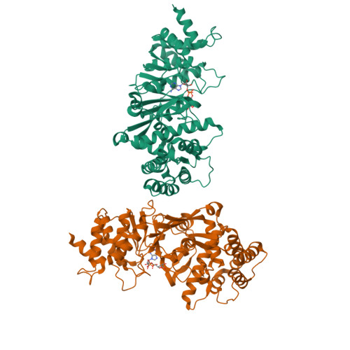
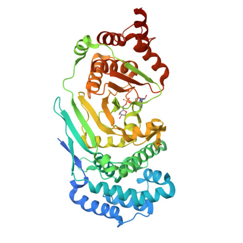
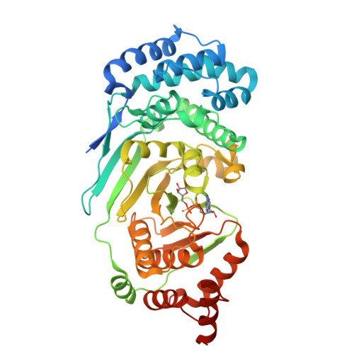
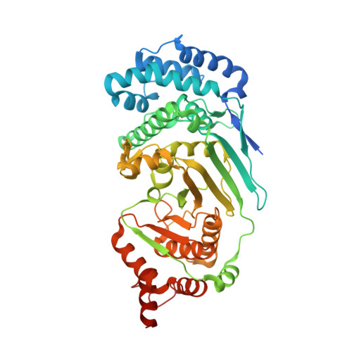
Structure and mechanism of a canonical poly(ADP-ribose) glycohydrolase.

Dunstan, M.S.Barkauskaite, E.Lafite, P.Knezevic, C.E.Brassington, A.Ahel, M.Hergenrother, P.J.Leys, D.Ahel, I.

(2012) Nat Commun 3: 878-878

  • DOI: https://doi.org/10.1038/ncomms1889
  • Primary Citation of Related Structures:  
    4EPP, 4EPQ

  • PubMed Abstract: 

    Poly(ADP-ribosyl)ation is a reversible post-translational protein modification involved in the regulation of a number of cellular processes including DNA repair, chromatin structure, mitosis, transcription, checkpoint activation, apoptosis and asexual development. The reversion of poly(ADP-ribosyl)ation is catalysed by poly(ADP-ribose) (PAR) glycohydrolase (PARG), which specifically targets the unique PAR (1''-2') ribose-ribose bonds. Here we report the structure and mechanism of the first canonical PARG from the protozoan Tetrahymena thermophila. In addition, we reveal the structure of T. thermophila PARG in a complex with a novel rhodanine-containing mammalian PARG inhibitor RBPI-3. Our data demonstrate that the protozoan PARG represents a good model for human PARG and is therefore likely to prove useful in guiding structure-based discovery of new classes of PARG inhibitors.


  • Organizational Affiliation

    Manchester Interdisciplinary Biocentre, Princess Street 131, M1 7DN, Manchester, UK.


Macromolecules
Find similar proteins by:  (by identity cutoff)  |  3D Structure
Entity ID: 1
MoleculeChains Sequence LengthOrganismDetailsImage
Poly(ADP-ribose) glycohydrolase
A, B
477Tetrahymena thermophilaMutation(s): 0 
EC: 3.2.1.143
UniProt
Find proteins for I6L8L8 (Tetrahymena thermophila)
Explore I6L8L8 
Go to UniProtKB:  I6L8L8
Entity Groups  
Sequence Clusters30% Identity50% Identity70% Identity90% Identity95% Identity100% Identity
UniProt GroupI6L8L8
Sequence Annotations
Expand
  • Reference Sequence
Experimental Data & Validation

Experimental Data

  • Method: X-RAY DIFFRACTION
  • Resolution: 1.95 Å
  • R-Value Free:  0.207 (Depositor), 0.200 (DCC) 
  • R-Value Work:  0.167 (Depositor), 0.160 (DCC) 
  • R-Value Observed: 0.169 (Depositor) 
Space Group: P 41
Unit Cell:
Length ( Å )Angle ( ˚ )
a = 112.7α = 90
b = 112.7β = 90
c = 88.6γ = 90
Software Package:
Software NamePurpose
SOLVEphasing
PDB_EXTRACTdata extraction
ADSCdata collection
XDSdata reduction
XDSdata scaling
PHENIXrefinement

Structure Validation

View Full Validation Report


Ligand Structure Quality Assessment 

Created with Raphaël 2.3.0Worse 01 BetterLigand structure goodness of fit to experimental dataBest fitted APRClick on this verticalbar to view details

Entry History 

Deposition Data

Revision History  (Full details and data files)

  • Version 1.0: 2012-06-20
    Type: Initial release
  • Version 1.1: 2024-02-28
    Changes: Data collection, Database references, Derived calculations