4F79 | pdb_00004f79

The crystal structure of 6-phospho-beta-glucosidase mutant (E375Q) in complex with Salicin 6-phosphate


Experimental Data Snapshot

  • Method: X-RAY DIFFRACTION
  • Resolution: 2.54 Å
  • R-Value Free: 
    0.229 (Depositor), 0.230 (DCC) 
  • R-Value Work: 
    0.180 (Depositor), 0.180 (DCC) 
  • R-Value Observed: 
    0.183 (Depositor) 

Starting Model: experimental
View more details

wwPDB Validation   3D Report Full Report


Ligand Structure Quality Assessment 

Created with Raphaël 2.3.0Worse 01 BetterLigand structure goodness of fit to experimental dataBest fitted P53Click on this verticalbar to view details

This is version 2.1 of the entry. See complete history


Literature

GH1-family 6-P-beta-glucosidases from human microbiome lactic acid bacteria.

Michalska, K.Tan, K.Li, H.Hatzos-Skintges, C.Bearden, J.Babnigg, G.Joachimiak, A.

(2013) Acta Crystallogr D Biol Crystallogr 69: 451-463

  • DOI: https://doi.org/10.1107/S0907444912049608
  • Primary Citation of Related Structures:  
    3QOM, 4F66, 4F79, 4GPN, 4GZE

  • PubMed Abstract: 

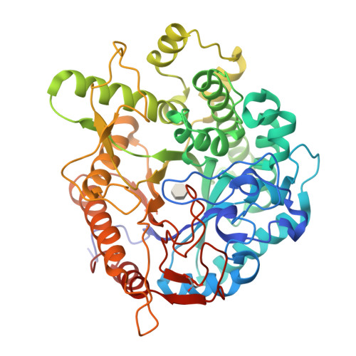
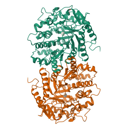
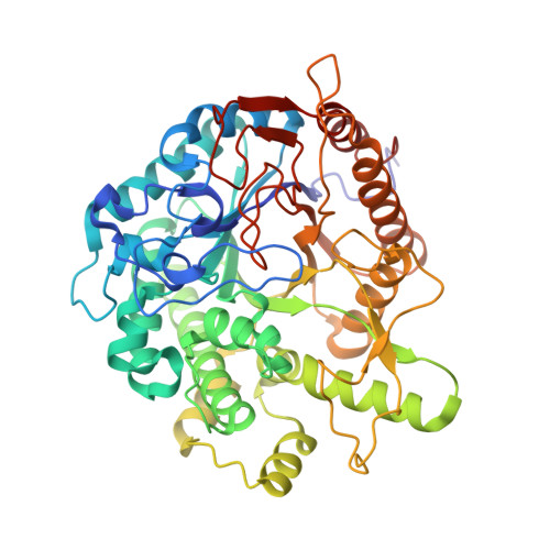
    In lactic acid bacteria and other bacteria, carbohydrate uptake is mostly governed by phosphoenolpyruvate-dependent phosphotransferase systems (PTSs). PTS-dependent translocation through the cell membrane is coupled with phosphorylation of the incoming sugar. After translocation through the bacterial membrane, the β-glycosidic bond in 6'-P-β-glucoside is cleaved, releasing 6-P-β-glucose and the respective aglycon. This reaction is catalyzed by 6-P-β-glucosidases, which belong to two glycoside hydrolase (GH) families: GH1 and GH4. Here, the high-resolution crystal structures of GH1 6-P-β-glucosidases from Lactobacillus plantarum (LpPbg1) and Streptococcus mutans (SmBgl) and their complexes with ligands are reported. Both enzymes show hydrolytic activity towards 6'-P-β-glucosides. The LpPbg1 structure has been determined in an apo form as well as in a complex with phosphate and a glucose molecule corresponding to the aglycon molecule. The S. mutans homolog contains a sulfate ion in the phosphate-dedicated subcavity. SmBgl was also crystallized in the presence of the reaction product 6-P-β-glucose. For a mutated variant of the S. mutans enzyme (E375Q), the structure of a 6'-P-salicin complex has also been determined. The presence of natural ligands enabled the definition of the structural elements that are responsible for substrate recognition during catalysis.


  • Organizational Affiliation

    Midwest Center for Structural Genomics, Biosciences Division, Argonne National Laboratory, Argonne, Illinois, USA.


Macromolecules
Find similar proteins by:  (by identity cutoff)  |  3D Structure
Entity ID: 1
MoleculeChains Sequence LengthOrganismDetailsImage
Putative phospho-beta-glucosidase480Streptococcus mutansMutation(s): 1 
Gene Names: bglSMU_1601
EC: 3.2.1.86
UniProt
Find proteins for Q8DT00 (Streptococcus mutans serotype c (strain ATCC 700610 / UA159))
Explore Q8DT00 
Go to UniProtKB:  Q8DT00
Entity Groups  
Sequence Clusters30% Identity50% Identity70% Identity90% Identity95% Identity100% Identity
UniProt GroupQ8DT00
Sequence Annotations
Expand
  • Reference Sequence
Experimental Data & Validation

Experimental Data

  • Method: X-RAY DIFFRACTION
  • Resolution: 2.54 Å
  • R-Value Free:  0.229 (Depositor), 0.230 (DCC) 
  • R-Value Work:  0.180 (Depositor), 0.180 (DCC) 
  • R-Value Observed: 0.183 (Depositor) 
Space Group: P 41 21 2
Unit Cell:
Length ( Å )Angle ( ˚ )
a = 82.278α = 90
b = 82.278β = 90
c = 221.211γ = 90
Software Package:
Software NamePurpose
SBC-Collectdata collection
MOLREPphasing
PHENIXrefinement
HKL-3000data reduction
HKL-3000data scaling

Structure Validation

View Full Validation Report


Ligand Structure Quality Assessment 

Created with Raphaël 2.3.0Worse 01 BetterLigand structure goodness of fit to experimental dataBest fitted P53Click on this verticalbar to view details

Entry History 

Deposition Data

Revision History  (Full details and data files)

  • Version 1.0: 2012-06-13
    Type: Initial release
  • Version 1.1: 2017-11-15
    Changes: Database references
  • Version 2.0: 2020-07-29
    Type: Remediation
    Reason: Carbohydrate remediation
    Changes: Atomic model, Data collection, Database references, Derived calculations
  • Version 2.1: 2023-09-13
    Changes: Data collection, Database references, Refinement description, Structure summary